
The Nervous System - Design: parts of the nervous system
The nervous system is a collection of cells, tissues, and organs. It can be split into two separate divisions: the central nervous system and the peripheral nervous system.
The central nervous system (CNS) acts as the command center of the body. It interprets incoming sensory information, then sends out instructions on how the body should react. The CNS consists of two major parts: the brain and the spinal cord.
The peripheral nervous system (PNS) is the part of the nervous system outside of the CNS. It consists mainly of nerves that extend from the brain and spinal cord to areas in the rest of the body. Cranial nerves carry impulses to and from the brain while spinal nerves carry impulses to and from the spinal cord. The PNS can be divided into two systems: the somatic nervous system and the autonomic nervous system. The somatic nervous system controls the voluntary movements of the skeletal muscles. The autonomic nervous system control activities in the body that are involuntary or automatic. These include the actions of the heart, glands, and digestive organs and associated parts.
The autonomic nervous system can be divided further into two subdivisions: the parasympathetic and sympathetic nervous systems. These two subdivisions work against each other. The parasympathetic nervous system regulates involuntary activities that keep the body running smoothly under normal, everyday conditions. The sympathetic nervous system controls involuntary activities that help the body respond to stressful situations.
- Arachnoid (ah-RAK-noid):
- Weblike middle layer of the three meninges covering the brain and spinal cord.
- Autonomic nervous system (aw-toh-NOM-ik NERV-us SIS-tem):
- Part of the peripheral nervous system that controls involuntary actions, such as the heartbeat, gland secretions, and digestion.
- Axon (AK-son):
- Taillike projection extending out a neuron that carries impulses away from the cell body.
- Basal ganglia (BAY-zul GANG-lee-ah):
- Paired masses of gray matter within the white matter of the cerebrum that help coordinate subconscious skeletal muscular movement.
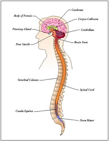
- Brain:
- Central controlling and coordinating organ of the nervous system.
- Cauda equina (KAW-da ee-KWHY-nah):
- Spinal nerves that hang below the end of the spinal cord.
- Central nervous system:
- Part of the nervous system consisting of the brain and spinal cord.
- Cerebral cortex (se-REE-bral KOR-tex):
- Outermost layer of the cerebrum made entirely of gray matter.
- Cerebrum (se-REE-brum):
- Largest part of the brain, involved with conscious perception, voluntary actions, memory, thought, and personality.
- Corpus callosum (KOR-pus ka-LOW-sum):
- Large band of neurons connecting the two cerebral hemispheres.
- Dendrites (DEN-drites):
- Branchlike extensions of neurons that carry impulses toward the cell body.
- Diencephalon (die-en-SEF-ah-lon):
- Rear part of the forebrain that connects the midbrain to the cerebrum and that contains the thalamus and hypothalamus.
- Dura mater (DUR-ah MAY-tur):
- Outermost and toughest of the three meninges covering the brain and spinal cord.
- Ganglion (GANG-glee-on):
- Any collection of nerve cell bodies forming a nerve center in the peripheral nervous system.
- Gray matter:
- Grayish nerve tissue of the central nervous system containing neuron cell bodies, neuroglia, and unmyelinated axons.
- Gyri (JYE-rye):
- Outward folds on the surface of the cerebral cortex.
- Hippocampus (hip-ah-CAM-pes):
- Structure in the limbic system necessary for the formation of long-term memory.
- Hypothalamus (hi-po-THAL-ah-mus):
- Region of the brain containing many control centers for body functions and emotions; also regulates the pituitary gland's secretions.
- Limbic system (LIM-bik SIS-tem):
- Group of structures in the cerebrum and diencephalon that are involved with emotional states and memory.
- Medulla oblongata (mi-DUL-ah ob-long-GAH-tah):
- Part of the brain located at the top end of the spinal cord that controls breathing and other involuntary functions.
- Meninges (meh-NIN-jeez):
- Membranes that cover the brain and spinal cord.
- Midbrain:
- Part of the brain between the hypothalamus and the pons that regulates visual, auditory, and rightening reflexes.
- Myelin (MY-ah-lin):
- Soft, white, fatty material that forms a sheath around the axons of most neurons.
- Nerve:
- Bundle of axons in the peripheral nervous system.
- Neuroglia (new-ROGUE-lee-ah):
- Also known as glial cells, cells that support and protect neurons in the central nervous system.
- Neuron (NUR-on):
- Nerve cell.
- Neurotransmitter (nur-oh-TRANS-mi-ter):
- Chemical released by the axon of a neuron that travels across a synapse and binds to receptors on the dendrites of other neurons or body cells.
- Node of Ranvier (NODE OF rahn-VEEAY):
- Small area between Schwann cells on an axon that is unmyelinated or uncovered.
- Oligodendrocyte (o-li-go-DEN-dro-site):
- Cell that produces the myelin sheath around the axons of neurons in the central nervous system.
- Parasympathetic nervous system (pair-ah-simpuh-THET-ik NERV-us SIS-tem):
- Division of the autonomic nervous system that controls involuntary activities that keep the body running smoothly under normal, everyday conditions.
- Peripheral nervous system (peh-RIFF-uh-ruhl NERV-us SIS-tem):
- Part of the nervous system consisting of the cranial and spinal nerves.
- Pia mater (PIE-ah MAY-tur):
- Delicate innermost layer of the three meninges covering the brain and spinal cord.
- Pons:
- Part of the brain connecting the medulla oblongata with the midbrain.
- Reflex (REE-flex):
- Involuntary and rapid response to a stimulus.
- Schwann cell (SHWAHN SELL):
- Cell that forms the myelin sheath around axons of neurons in the peripheral nervous system.
- Somatic nervous system (so-MAT-ik NERV-us SIS-tem):
- Part of the peripheral nervous system that controls the voluntary movements of the skeletal muscles.
- Spinal cord:
- Long cord of nerve tissue running through the spine or backbone that transmits impulses to and from the brain and controls some reflex actions.
- Sulci (SUL-sye):
- Shallow grooves on the surface of the cerebral cortex.
- Sympathetic nervous system (sim-puh-THET-ik NERV-us SIS-tem):
- Division of the autonomic nervous system that controls involuntary activities that help the body respond to stressful situations.
- Synapse (SIN-aps):
- Small space or gap where a nerve impulse passes between the axon of one neuron and a dendrite of the next neuron.
- Thalamus (THAL-ah-mus):
- Part of the brain behind the hypothalamus that acts as the brain's main relay station, sending information to the cerebral cortex and other parts of the brain.
- White matter:
- Whitish nerve tissue of the central nervous system containing bundles of myelinated axons.
Neurons
The cells making up the brain, spinal cord, and nerves are called neurons. They are special cells capable of receiving a stimulus (nerve or electrical impulse), transmitting that stimulus throughout their length, and then delivering that stimulus to other cells next to them. The human body contains about 200 billion neurons. Almost half of them are located in the brain.
A neuron consists of three main parts: the cell body, dendrites, and an axon (dendrites and axons are both referred to as nerve fibers). The cell body has most of the same structures found in typical body cells, such as a nucleus (the part of the cell that controls its activities). It is ball shaped, about 0.001 inch (0.002 centimeter) in diameter.
Dendrite comes from the Greek word dendron , meaning "tree." Dendrites are hairlike threads branching off of the cell body like branches of a tree. Extensions of the cell body, they contain the same cytoplasm or cellular fluid found in the cell body. Dendrites are the points through which signals from adjacent neurons enter a particular neuron (the signal is then transmitted to the cell body). Since each neuron contains many dendrites, a neuron can receive signals from many other surrounding neurons.
An axon is a taillike projection extending out of one end of the cell body. It ends in a cluster of branches called terminal branches or axon terminals. Axons have the opposite function of dendrites: they carry nerve impulses away from the cell body. Axons vary in length and diameter. Some (such as those in the central nervous system) are very short, no longer than 0.01 inch (0.02 centimeter). Others (such as those in the peripheral nervous system) can be 3 feet (1 meter) long.
Most long axons are surrounded by a white, fatty material called myelin. The tubelike covering formed is known as a myelin sheath. It serves the same kind of function as the wrapping on a telephone line or an electrical cable. It protects the axon and prevents electrical impulses traveling through it from becoming lost.
Special cells form the myelin sheath by wrapping themselves around the axons of neurons. In the CNS, the cells forming the myelin sheath are called oligodendrocytes. In the PNS, special cells known as Schwann cells form the myelin sheath. The gap or indentation on an axon where one Schwann cell ends and another begins is known as a node of Ranvier. The nodes are unmyelinated (lack a myelin sheath), and the nerve or electrical impulse jumps from node to node as it passes along an axon (in unmyelinated axons, the impulse travels continuously along the axon).
Scientists believe Schwann cells produce a chemical that helps regenerate or restore damaged neurons in the peripheral nervous system. For example, if surgeons are able to reattach a person's severed hand, that person may regain some sensation and movement in that hand as neurons grow and make connections. Conversely, oligodendrocytes lack this ability. This is why an injury to the brain or spinal cord often results in some permanent loss of function.
TYPES OF NEURONS. Neurons in the body may be divided into three groups: sensory neurons, motor neurons, and interneurons. As their name implies, sensory neurons carry impulses or sensations from receptors to the brain or spinal cord (central nervous system). Receptors, which are located in the skin, skeletal muscles, joints, and internal organs, detect changes both inside and outside the body. Motor neurons work in the opposite direction. They carry impulses from the brain or spinal cord to muscles and glands, causing muscles to contract and glands to secrete. Both sensory and motor neurons make up the peripheral nervous system. Interneurons work entirely within the central nervous system. They conduct impulses from sensory to motor neurons.

American neuroendocrinologist Susan E. Leeman (1930– ) is known for her work with substance P, a peptide that helps govern the functioning of the nervous and lymphatic systems. (A peptide is a compound containing two or more amino acids, the building blocks of proteins.) While doing research on protein-hormones, Leeman found a peptide that could stimulate the secretion of saliva. The chemical turned out to be substance P, which had been discovered in the 1930s but had never been isolated (separated from other substances for individual study).
Leeman and her colleagues, working at Brandeis University in Massachusetts, isolated and characterized the peptide. A nerve transmitter that has many functions in the body, substance P is distributed throughout both the central and peripheral nervous systems. Substance P is important in the interaction between the nervous system and the lymphatic system (which governs body immunity) and seems to play a role in inflammation in the body.
Each neuron carries impulses in only one direction. This prevents impulses from traveling both ways in a neuron and canceling each other when they meet.
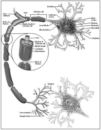
SUPPORTING CELLS. Neuroglia, or glial cells, are cells that surround neurons in the central nervous system. They do not conduct impulses, but help to support and protect neurons, combining with them to form what is known as nerve tissue. They also supply neurons with nutrients and remove their wastes. Neuroglia are abundant, accounting for some ten times the number of neurons. An example of neuroglia in the CNS are oligodendrocytes.
In the PNS, neurons are supported by Schwann cells and satellite cells (which form around the cell body to protect and cushion it).
Nerves
A nerve is a bundle of axons in the PNS. Each axon or nerve fiber is wrapped in delicate connective tissue. Groups of axons are then bound in coarser connective tissue to form bundles. Finally, many bundles are bound together (along with blood vessels to nourish the axons and Schwann cells) by even tougher connective tissue to form a nerve.
Nerves are categorized like neurons according to the direction in which they conduct impulses. Sensory nerves, made of the axons of sensory neurons, carry impulses to the brain and spinal cord. Motor nerves, made of the axons of motor neurons, carry impulses to the muscles and glands. Mixed nerves contain axons of both sensory and motor neurons. The most abundant nerves, mixed nerves can conduct impulses both to and from the central nervous system.
The brain
The human brain is a soft, shiny, grayish white, mushroom-shaped structure encased within the skull. At birth, a typical human brain weighs between 12 and 14 ounces (350 and 400 grams). By the time an average person reaches adulthood, the brain weighs about 3 pounds (1.36 kilograms). Because of greater average body size, the brains of male are generally about 10 percent larger than those of females. Although brain size varies considerably among humans, there is no correlation or link between brain size and intelligence.
The human brain is composed of up to one trillion nerve cells. One hundred billion of these are neurons, and the remainder are the supporting neuroglia. The brain consists of gray and white matter. Gray matter is nerve tissue in the CNS composed of neuron cell bodies, neuroglia, and unmyelinated axons; white matter is nerve tissue in the CNS composed chiefly of bundles of myelinated axons.
The brain is protected by the skull and by three membranes called the meninges. The outermost membrane is known as the dura mater, the middle as the arachnoid, and the innermost as the pia mater. Also protecting the brain is cerebrospinal fluid, a liquid that circulates between the arachnoid
and pia mater. Many arteries and veins on the surface of the brain penetrate inward. Glucose, oxygen, and certain ions pass easily from the blood into the brain; other substances, such as antibiotics, do not. Scientists believe capillary walls create a blood-brain barrier that protects the brain from a number of biochemicals circulating in the blood.
The parts of the brain can be divided in terms of structure and function. The four principal sections of the human brain are the brain stem, the diencephalon, the cerebrum, and the cerebellum.
THE BRAIN STEM. The brain stem is the stalk of the brain and is a continuation of the spinal cord. It consists of the medulla oblongata, pons, and midbrain. The medulla oblongata is actually a portion of the spinal cord that extends into the brain. All messages that are transmitted between the brain and spinal cord pass through the medulla. Nerves on the right side of the medulla cross to the left side of the brain, and those on the left cross to the right. The result of this arrangement is that each side of the brain controls the opposite side of the body.
Three vital centers in the medulla control heartbeat, rate of breathing, and diameter of the blood vessels. Centers that help coordinate swallowing, vomiting, hiccuping, coughing, sneezing, and other basic functions of life are also located in the medulla. A region within the medulla helps to maintain the conscious state. The pons (from the Latin word meaning "bridge") conducts messages between the spinal cord and the rest of the brain, and between the different parts of the brain. The midbrain conveys impulses from the hypothalamus to the pons and spinal cord. It also contains visual and audio reflex centers involving the movement of the eyeballs and head.
Twelve pair of cranial nerves originate in the underside of the brain, mostly from the brain stem. They leave the skull through openings and extend as peripheral nerves to their destinations. Cranial nerves bring information to the brain from regions in the face, head, and neck. For example, the olfactory nerve transmits messages about smell from the nose and the optic nerve transmits visual information from the eyes. The only exception is the vagus nerve (vagus comes from the Latin word meaning "wandering"). It is the lone cranial nerve that serves other areas of the body. The vagus nerve branches extensively to the larynx, heart, lungs, stomach, and intestines. Among other functions, it helps promote digestive activity and regulate heart activity.
THE DIECEPHALON. The diencephalon lies above the brain stem, and includes the thalamus and hypothalamus. The thalamus is an important relay station for sensory information coming to the cerebral cortex from other parts of the brain. The thalamus also interprets sensations of pain, pressure, temperature, and touch, and is concerned with some of our emotions and memory. It receives information from the outside environment in the form of sound, smell, and taste.
The hypothalamus performs numerous important functions. These include the control of the autonomic nervous system. The hypothalamus controls normal body temperature and helps regulate the endocrine system, which produces hormones or chemical messengers that regulate body functions (for a further discussion of these actions, see chapter 3). It informs the body when it is hungry, full, or thirsty. It helps regulate sleep and wakefulness and is involved in the emotions of anger and aggression.
THE CEREBRUM. The cerebrum makes up about 80 percent of the brain's weight. It lies above the diencephalon. The cerebrum's outer layer, the cerebral cortex, is made entirely of gray matter (white matter makes up the inner portion of the cerebrum). The tissue of the cerebral cortex is about 0.08 to 0.16 inch (2 to 4 millimeters) thick. The cerebral cortex is folded extensively. The folds are called convolutions or gyri, and the shallow grooves between the folds are sulci. Deeper grooves, which are less numerous, are called fissures. The folds greatly increase the surface area of the cerebral cortex—it would have a surface area of about 5 square feet (1.5 square meters) if spread out—and thus the total number of nerve cell bodies it contains.
Located deep within the white matter of the cerebrum just above the diencephalon are two paired masses of gray matter known as basal ganglia. They are important in coordinating subconscious skeletal muscular movement, such as swinging of the arms while walking.
A deep fissure separates the cerebrum into a left and right hemisphere or half. The corpus callosum, a bundle of more than 200 million neurons, connects the two cerebral hemispheres and carries vast amounts of information between them—an estimated 4 billion nerve impulses per second. By studying patients whose corpus callosum had been destroyed, scientists have learned that differences exist between the left and right hemispheres. The left side of the brain functions mainly in speech, logic, writing, and arithmetic. The right side of the brain, on the other hand, is more concerned with imagination, art, symbols, and spatial relations.
Sperm whale: 17 pounds (7.8 kilograms)
Elephant: 13.2 pounds (6 kilograms)
Bottle-nosed dolphin: 3.3 pounds (1.5 kilograms)
Human (adult): 3 pounds (1.36 kilograms)
Camel: 1.5 pounds (0.76 kilogram)
Hippopotamus: 1.3 pounds (0.58 kilogram)
Polar bear: 1.1 pounds (0.5 kilogram)
Chimpanzee: 14.7 ounces (420 grams)
Lion: 8.4 ounces (240 grams)
Dog: 2.5 ounces (72 grams)
Cat: 1.1 ounces (30 grams)
Rabbit: 0.4 ounce (11.5 grams)
Squirrel: 0.26 ounce (7.6 grams)
Hamster: 0.05 ounce (1.4 grams)
Bull frog: 0.008 ounce (0.24 gram)
Scientists have further divided each cerebral hemisphere into lobes named after the overlying bones of the skull: frontal (forehead area), temporal (on the sides above the ears), parietal (top part of the head), and occipital (back of the head) lobes.
The cerebral cortex is the portion of the brain that provides the most important distinctions between humans and other animals. It is responsible for the vast majority of functions that define what is meant by "being human." It enables humans not only to receive and interpret all kinds of sensory information, such as color, odor, taste, and sound, but also to remember, analyze, interpret, make decisions, and perform a host of other "higher" brain functions.
By studying animals and humans who have suffered damage to the cerebral cortex, scientists have found that the various lobes house areas with specific functions. The frontal lobes contain motor areas that generate impulses for voluntary movements. An area usually located in the left frontal lobe is called Broca's area. It coordinates the movements of the mouth involved in speaking. The parietal lobes contain general sensory areas that receive impulses from receptors in the skin. The temporal lobes contain auditory areas (receive impulses from the ears for hearing) and olfactory areas (receive impulses from receptors in the nose for smell). The occipital lobes contain visual areas that receive impulses from the retinas of the eyes. Different areas in the occipital lobes are concerned with judging distance and other spatial relationships.
German-born American theoretical physicist Albert Einstein (1879–1955), who formulated the theory of relativity (an approach for studying the nature of the universe), is considered by many to have been one of the greatest physicists of all time.
When Einstein died in 1955, the physician who performed the autopsy removed his brain from his body in order to perform scientific studies on it. For years, however, the brain was kept in a jar (for a while, the jar was even placed in a cardboard box behind a beer cooler). Although the physician took measurements of Einstein's brain and cut it into 240 pieces of varying size, he published none of his findings.
In 1996, the physician allowed Canadian researchers to study the remains of Einstein's brain. What the researchers found might explain the reason Einstein was a genius. They discovered that a region in Einstein's brain was 15 percent larger than the same area in people with average intelligence. That region controls mathematical thought, spatial relationships, and other mental processes. Known as the inferior parietal lobe, it is located about the level of the ear, starting in the front of the brain and extending twothirds of the way back.
The researchers believe the enlarged region created a space for more neurons to make connections between each other and to work together more easily.
Association areas, those not involved with a particular movement or sensation, are located in all the lobes. These areas are concerned with emotions and intellectual processes. In association areas, innumerable impulses are processed that result in memory, emotions, judgment, personality, and intelligence: what truly makes each person an individual.
THE CEREBELLUM. The cerebellum is located below the cerebrum and behind the brain stem, and is shaped like a butterfly. The "wings" are the cerebellar hemispheres, and each consists of lobes that have distinct grooves or fissures. The cerebellum controls the actions of the muscular system needed for movement, balance, and posture. All motor activity in the body depends on the cerebellum.
THE LIMBIC SYSTEM. The limbic system is a horseshoe-shaped area of the brain located along the border between the cerebrum and diencephalon. Key structures of the limbic system include the almond-shaped amygdala and the sea horse-shaped hippocampus. The limbic system is concerned with emotional states (such as rage, fear, and sexual arousal) and memory. The hippocampus, in particular, plays a vital role in learning and long-term memory.
The spinal cord
The spinal cord, a glistening white rope, is a continuation of the brain stem. It transmits impulses to and from the brain and controls some reflex actions. On average, the spinal cord measures about 18 inches (45 centimeters) in length and about 0.5 inch (14 centimeters) in width. It weighs about 1.25 ounces (35 grams).
The vertebral column or backbone encloses the spinal cord. This long channel, made of individual bones called vertebrae, protects the spinal cord from mechanical injury. Like the brain, the spinal cord is also cushioned and protected by meninges. Arteries run along the surface of the spinal cord, supplying it with a nourishing blood supply.
The spinal cord is composed of roughly 13.5 million neurons. It appears white because its thick outer layer is made of white matter. This layer contains many myelinated axons that form bundles—called tracts—that carry either sensory information or motor commands. Tracts that carry sensory information toward the brain are called ascending tracts. Those that carry motor commands from the brain into the spinal cord are called descending tracts.
Within the spinal cord is an H- or butterfly-shaped gray area composed of gray matter. Cell bodies of neurons and supporting neuroglia make up this gray matter. Many of these cell bodies are those of motor neurons. Their axons pass out of the spinal cord to control skeletal muscles or to regulate the actions of smooth muscles, cardiac muscles, and glands.
Extending out from the spinal cord between the vertebrae are thirty-one pair of spinal nerves. All these nerves are mixed nerves, containing thousands of axons of both sensory and motor neurons. Inside the vertebral column, however, each nerve is split into two branches that connect with the spinal cord. The branch that attaches on the rear (posterior) portion of the spinal cord is called the dorsal root. It contains the axons of sensory neurons. On
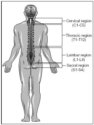
each dorsal root is an enlarged area called the dorsal root ganglion (a ganglion is any collection of neuron cell bodies in the PNS). This ganglion contains the cell bodies of the sensory neurons. The branch that attaches on the front (anterior) portion of the spinal cord is called the ventral root. It contains the axons of motor neurons.
The thirty-one pairs of spinal nerves exit the vertebral column to serve the areas of the body close by. The first or top eight pairs (located in the neck area) bring impulses to and from the head, neck, shoulders, arms, and diaphragm. The next twelve pairs (located in the chest area) bring impulses to and from the trunk of the body, including internal organs such as the heart and lungs. The remaining eleven pairs bring impulses to and from the lower part of the body—the hips, pelvic cavity, and legs. Damage to a spinal nerve or either of its roots will result in the loss of sensation and in paralysis of the area of the body being served by that nerve.
The spinal cord does not extend all the way down the vertebral column. In order to reach their proper openings to exit the column, the last eleven pair of spinal nerves hang below the end of the spinal cord like long hairs. Because of their appearance, they are collectively called the cauda equina (in Latin, cauda equina means "horse's tail").