
The Integumentary System - Design: parts of the integumentary system
Integument comes from the Latin word integumentum , meaning "cover" or "enclosure." In animals and plants, an integument is any natural outer covering, such as skin, shell, membrane, or husk. The human integumentary system is an external body covering, but also much more. It protects, nourishes, insulates, and cushions. It is absolutely essential to life. Without it, an individual would be attacked immediately by bacteria and die from heat and water loss.
The integumentary system is composed primarily of the skin and accessory structures. Those structures include hair, nails, and certain exocrine glands (glands that have ducts or tubes that carry their secretions to the surface of the skin or into body cavities for elimination).
Skin
Although the skin is not often thought of as an organ, such as the heart or liver, medically it is. An organ is any part of the body formed of two or more tissues that performs a specialized function. As an organ, the skin is the largest and heaviest in the body. In an average adult, the skin covers about 21.5 square feet (2 square meters) and accounts for approximately 7 percent of body weight, or about 11 pounds (5 kilograms) in a 160-pound (73-kilogram) person. It ranges in thickness from 0.04 to 0.08 inches (1 to 2 millimeters), but can measure up to 0.2 inches (6 millimeters) thick on the palms of the hands and the soles of the feet. The skin in these areas is referred to as thick skin (skin elsewhere on the body is called thin skin).
- Apocrine sweat glands (AP-oh-krin):
- Sweat glands located primarily in the armpit and genital areas.
- Arrector pili muscle (ah-REK-tor PI-li):
- Smooth muscle attached to a hair follicle that, when stimulated, pulls on the follicle, causing the hair shaft to stand upright.
- Dermal papillae (DER-mal pah-PILL-ee):
- Finger-like projections extending upward from the dermis containing blood capillaries, which provide nutrients for the lower layer of the epidermis; also form the characteristic ridges on the skin surface of the hands (fingerprints) and feet.
- Dermis (DER-miss):
- Thick, inner layer of the skin.
- Eccrine sweat glands (ECK-rin):
- Body's most numerous sweat glands, which produce watery sweat to maintain normal body temperature.
- Epidermis (ep-i-DER-miss):
- Thin, outer layer of the skin.
- Epithelial tissue (ep-i-THEE-lee-al):
- Tissue that covers the internal and external surfaces of the body and also forms glandular organs.
- Integument (in-TEG-ye-ment):
- In animals and plants, any natural outer covering, such as skin, shell, membrane, or husk.
- Keratin (KER-ah-tin):
- Tough, fibrous, water-resistant protein that forms the outer layers of hair, calluses, and nails and coats the surface of the skin.
- Lunula (LOO-noo-la):
- White, crescent-shaped area of the nail bed near the nail root.
- Melanocyte (MEL-ah-no-site):
- Cell found in the lower epidermis that produces the protein pigment melanin.
- Organ (OR-gan):
- Any part of the body formed of two or more tissues that performs a specialized function.
- Sebaceous gland (suh-BAY-shus):
- Exocrine gland in the dermis that produces sebum.
- Sebum (SEE-bum):
- Mixture of oily substances and fragmented cells secreted by sebaceous glands.
- Squamous cells (SKWA-mus):
- Cells that are flat and scalelike.
- Subcutaneous (sub-kew-TAY-nee-us):
- Tissues between the dermis and the muscles.
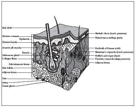
The skin has two principal layers: the epidermis and the dermis. The epidermis is the thin, outer layer, and the dermis is the thicker, inner layer. Beneath the dermis lies the subcutaneous layer or hypodermis, which is composed of adipose or fatty tissue. Although not technically part of the skin, it does anchor the skin to the underlying muscles. It also contains the major blood vessels that supply the dermis and houses many white blood cells, which destroy foreign invaders that have entered the body through breaks in the skin.
EPIDERMIS. The epidermis is made of stratified squamous epithelial tissue. Epithelial tissue covers the internal and external surfaces of the body and also forms glandular organs. Squamous cells are thin and flat like fish scales. Stratified simply means having two or more layers. In short, the epidermis is composed of many layers of thin, flattened cells that fit closely together and are able to withstand a good deal of abuse or friction.
The epidermis can be divided into four or five layers. Most important of these are the inner and outer layers. The inner or deepest cell layer is the only layer of the epidermis that receives nutrients (from the underlying dermis). The cells of this layer, called basal cells, are constantly dividing and creating new cells daily, which push the older cells toward the surface. Basal cells produce keratin, an extremely durable and water-resistant fibrous protein.
Another type of cell found in the lower epidermis is the melanocyte. Melanocytes produce melanin, a protein pigment that ranges in color from yellow to brown to black. The amount of melanin produced determines skin color, which is a hereditary characteristic. The melanocytes of dark-skinned individuals continuously produce large amounts of melanin. Those of light-skinned individuals produce less. Freckles are the result of melanin clumping in one spot.
The outermost layer of the epidermis consists of about twenty to thirty rows of tightly joined flat dead cells. All that is left in these cells is their keratin, which makes this outer layer waterproof. It takes roughly fourteen days for cells to move from the inner layer of the epidermis to the outer layer. Once part of the outer layer, the dead cells remain for another fourteen days or so before flaking off slowly and steadily.
DERMIS. The dermis, the second layer of skin, lies between the epidermis and the subcutaneous layer. Much thicker than the epidermis, the dermis contains the accessory skin structures. Hair, sweat glands, and sebaceous (oil) glands are all rooted in the dermis. This layer also contains blood vessels and nerve fibers. Nourished by the blood and oxygen provided by these blood vessels, the cells of the dermis are alive.
Connective tissue forms the dermis. Bundles of elastic and collagen (tough fibrous protein) fibers blend into the connective tissue. These fibers provide the dermis strength and flexibility.
The upper layer of the dermis has fingerlike projections that extend into the epidermis. Called dermal papillae, they contain blood capillaries that provide nutrients for the basal cells in the epidermis. On the skin surface of the hands and feet, especially on the tips of the fingers, thumbs, and toes, the dermal papillae form looped and whorled ridges. These print patterns, known as fingerprints or toeprints, increase the gripping ability of the hands and feet. Genetically determined, the patterns are unique to every individual.
Fingerprints (the pattern of ridges on an individual's fingertips and thumbs formed by dermal papillae) are unique to each individual and the patterns never change. People have long known about the distinctiveness of fingerprints, but their use in identifying people did not arise until the nineteenth century.
It is generally acknowledged that English scientist Francis Galton (1822–1911) was the first person to devise a system of fingerprint identification. In the 1880s, Galton obtained the first extensive collection of fingerprints for his studies on heredity. He also established a bureau for the registration of civilians by means of fingerprints and measurements.
Galton's ideas were further developed by fellow Englishman Edward R. Henry (1850–1931). In the 1890s, Henry developed a more simplified fingerprint classification system. In 1901, he established England's first fingerprint bureau, called the Fingerprint Branch, within the Scotland Yard police force. Henry's system is still used today in Great Britain and the United States.
Within the dermis are sensory receptors for the senses of touch, pressure, heat, cold, and pain. A specific type of receptor exists for each sensation. For pain, the receptors are free nerve endings. For the other sensations, the receptors are encapsulated nerve endings, meaning they have a cellular structure around their endings. The number and type of sensory receptors present in a particular area of skin determines how sensitive that area is to a particular sensation. For example, fingertips have many touch receptors and are quite sensitive. The skin of the upper arm is less sensitive because it has very few touch receptors.
Accessory structures
The accessory structures of the integumentary system include hair, nails, and sweat and sebaceous glands.
HAIR. Roughly 5 million hairs cover the body of an average individual. About 100,000 of those hairs appear on the scalp. Almost every part of the body is covered by hair, except the palms of the hands, the soles of the feet, the sides of the fingers and toes, the lips, and certain parts of the outer genital organs.
Each hair originates from a tiny tubelike structure called a hair follicle that extends deep into the dermis layer. Often, the follicle will project into the subcutaneous layer. Capillaries and nerves attach to the base of the follicle, providing nutrients and sensory information. Inside the base of the follicle, epithelial cells grow and divide, forming the hair bulb or enlarged hair base. Keratin, the primary component in these epithelial cells, coats and stiffens the hair as it grows upward through the follicle. The part of the hair enclosed in the follicle is called the hair root. Once the hair projects from the scalp or skin, it is called a hair shaft.
The older epithelial cells forming the hair root and hair shaft die as they are pushed upward from the nutrient-rich follicle base by newly formed cells. Like the upper layers of the epidermis, the hair shaft is made of dead material, almost entirely protein. The hair shaft is divided into two layers: the cuticle or outer layer consists of a single layer of flat, overlapping cells; the cortex or inner layer is made mostly of keratin.
Hair shafts differ in size, shape, and color. In the eyebrows, they are short and stiff, but on the scalp they are longer and more flexible. Elsewhere on the body they are nearly invisible. Oval-shaped hair shafts produce wavy hair. Flat or ribbonlike hair shafts produce kinky or curly hair. Perfectly round hair shafts produce straight hair. The different types of melanin—yellow, rust, brown, and black—produced by melanocytes at the follicle base combine to create the many varieties of hair color, from the palest blonde to the richest black. With age, the production of melanin decreases, and hair color turns gray.
Attached to each hair follicle is a ribbon of smooth muscle called an arrector pili muscle. When stimulated, the muscle contracts and pulls on the follicle, causing the hair shaft to stand upright.
NAILS. Nails in humans correspond to the hooves of horses and cattle and the claws of birds and reptiles. Found on the ends of fingers and toes, nails are produced by nail follicles just as hair is produced by hair follicles. The nail root is that portion of the nail embedded in the skin, lying very near the bone of the fingertip. Here, cells produce a stronger form of keratin than is found in hair. As new cells are formed, older cells are pushed forward, forming the nail body or the visible attached portion of the nail. The free edge is that portion of the nail that extends over the tip of the finger or toe. Healthy fingernails grow about 0.04 inches (1 millimeter) per week, slightly faster than toenails.
The nail body is made of dead cells, but the nail bed (the tissue underneath the nail body) is alive. The blood vessels running through the nail bed give the otherwise transparent nail body a pink color. Near the nail root, however, these blood vessels are obscured. The resulting white crescent is called the lunula (from the Latin word luna , meaning "moon").
SWEAT GLANDS. More than 2.5 million sweat glands are distributed over most surfaces of the human body. They are divided into two types: eccrine sweat glands and apocrine sweat glands.
Eccrine glands, the more numerous of the two types, are found all over the body. They are especially numerous on the forehead, upper lip, palms, and soles. The glands are simply coiled tubes that originate in the dermis. A duct extends from the gland to the skin's surface, where it opens into a pore. Eccrine glands produce sweat or perspiration, a clear secretion that is 99 percent water. Some salts, traces of waste materials such as urea, and vitamin C form the remainder (the salts give sweat its characteristic salty taste).
Depending on temperature and humidity, an average individual loses 0.6 to 1.7 quarts (0.3 to 0.8 liters) of water every day through sweating. During rigorous physical activity or on a hot day, that amount could rise to 5.3 to 7.4 quarts (5 to 7 liters).
Apocrine glands are found in the armpits, around the nipples, and in the groin. Like eccrine glands, apocrine glands are coiled tubes found in the dermis. However, they are usually larger and their ducts empty into hair follicles. Also, apocrine glands do not function until puberty. At that time, they begin to release an odorless cloudy secretion that contains fatty acids and protein. If the secretion of apocrine glands is allowed to remain on the skin for any length of time, bacteria that lives on the skin breaks down the fatty acids and protein for their growth, creating the unpleasant odor often associated with sweat.
Apocrine glands are activated by nerve fibers during periods of pain and stress, but their function in humans is not well understood. Scientists theorize they may act as sexual attractants.
SEBACEOUS GLANDS. Sebaceous glands, also known as oil glands, are found in the dermis all over the body, except for the palms and soles. They secrete sebum, a mixture of lipids (fats), proteins, and fragments of dead fatproducing cells. The function of sebum is to prevent the drying of skin and hair. It also contains chemicals that kill bacteria present on the skin surface. While most sebaceous glands secrete sebum through ducts into hair follicles, some secrete sebum directly onto the surface of the skin. Arrector pili muscles, which contract to elevate hairs, also squeeze sebaceous glands, forcing out sebum.