
The Sense Organs - The eye
Rather than take a name-by-name anatomical tour of the eye, let's instead take just three programmed tours of the eye to explore:
- • the transformation of light energy into vision;
- • focusing, or how the structure of the eye prepares light for transformation into vision; and
- • the supporting structures and service units of the eye.
Transforming Light Energy into Vision
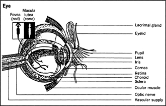
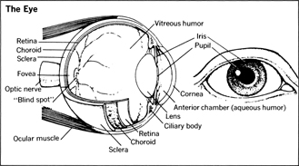
If you could look through the opening in the front of your eye (the pupil ), and see to the very back surface of your eye (as if you could see through the needle valve opening to the inside skin of a basketball), you would see your own retina . On your retina are located all the sense receptor cells that enable us to see. There are none anywhere else in the body.
Rods and Cones
A good argument can be made that man's vision is really two senses. For on the paper-thin retina are two quite anatomically distinct sense receptors (nerve endings) named, for their appearance under high magnification, cones and rods . The cones are concentrated at a tiny spot on the retina called the fovea .
If the focusing machinery of the eye (cornea, lens, etc.) is working just right, light rays from the outside have their sharpest focus on the fovea. Surrounding the fovea is a yellowish area called the macula lutea (Latin, “yellow spot”). Together, the fovea and macula lutea make a circle not much bigger than the head of a pin.
All our seeing of colors and fine details is accomplished by the cones of the fovea and the macula lutea. Beyond the yellowish circumference of the macula lutea, there are fewer and fewer cones: rods become the dominant structures on the retina. It has been estimated that there are something fewer than 10 million cones on the retina of each eye, but more than 10 times as many rods—100 million of them or more.
Although the tight circle of cones in each eye gives man his ability to do close, detailed work (including reading) and to discriminate colors, the cones are virtually useless in detecting objects that are a bit off center from our direct focus, and furthermore operate only in good lighting conditions or in response to bright light sources.
The rods compensate for the specialized limits of the cones. They take over completely in dim light, and also give us the ability to detect peripheral objects and movements—"out of the corner of the eye.” Because the rods are not sensitive to colors, our seeing at night is almost completely in black-and-white.
You can give yourself an interesting demonstration of the interacting functions of your own cones and rods if you walk from bright, sunny daylight into a dimly lit theater. In the bright light, your cones have been picking out sharp images and colors. But once in dimness, the cones become inoperative. For a few moments, in fact, you may see almost nothing at all.
Visual Purple
The momentary interval after the cones stop working but before the rods begin to function is explained by a curious pigment present in the eye called visual purple . This substance is manufactured constantly by the rods, and must be present for the rods to respond to dim light—but it is destroyed when exposed to bright light. Thus, after entering a darkened room, it takes a few moments for visual purple to build up in the retina. One of the principal constituents of visual purple is vitamin A, which is why this vitamin (present in carrots and other yellow produce) is said to increase our capacity to see in the dark.
The Optic Nerve
Every nerve ending is part of a larger unit, a neuron or nerve cell, and the sense receptors called rods and cones are no exception. Like all nerve cells, each rod and cone sports a long nerve fiber or axon leading away from the site of reception. In each eye, fibers serving the hundred-million-plus rods and cones all converge at a certain spot just behind the retina, forming the optic nerve . There are no rods and cones at the point where the optic nerve exits from behind the retina at the back of the eyeball: that is why everybody has a “blind spot” at that point. An image passing through that spot completely disappears.
From the retinas, both optic nerves set a course almost directly through the middle of the brain. Right and left optic nerves converge, their individual fibers partially intertwining a short distance behind the eyes. Then this joint optic nerve trunk proceeds toward the rear of the head, where the occipital lobes , the brain's “centers for seeing,” are located. Just before reaching the occipital lobes, the optic nerve splits again into thousands of smaller nerve bundles (called visual radiations ) that disappear into the visual cortex or “outer bark” of the occipital lobes. Only at this point are the bits of light energy that have stimulated our rods and cones transformed into images that our brain can “see.”
Focusing and Light Control
Lacking the structures of the retina and their connection to the brain via the optic nerve, we could not see. Lacking reasonably normal functioning of the cornea, lens, and iris, we do not see well.
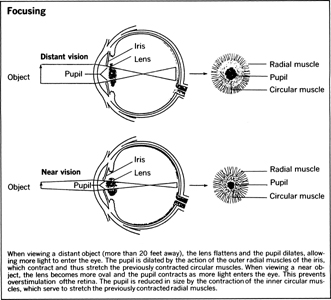
Good vision depends upon the eye being able to bend incoming light rays in such a way that the image being observed falls directly on the retina—in other words, upon proper focusing. The bending or refraction of light rays is the joint work of two curves, transparent slivers of specialized tissue through which light passes on its way to the retina. These are the cornea and the lens . Broadly speaking, the degree of curvature and thickness of these two structures determines whether we see well or poorly, are nearsighted or farsighted.
The Cornea and Lens
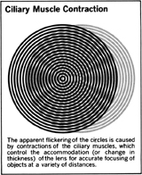
The cornea, which does the major light-bending, has a virtually fixed curvature and thickness. The lens puts the finishing touches on the focusing. Its thickness and curvature are adjustable—more or less without our conscious awareness—depending on whether we wish to focus on something nearer or farther away. The lens is made thicker or thinner, a process called accommodation , by the relaxing and contracting of tiny, attached ciliary muscles . Normally, these muscles do not have to work at all if we are looking at objects more than 20 feet away: but they often are overworked by a great deal of close work.
The Pupil and Iris
Effective focusing in various light conditions also depends upon the diameter of the hole through which light enters the eye. This hole is the pupil . Its diameter is controlled automatically—wider in dim light, narrower in bright light—by the muscles of the surrounding iris . The iris muscle contains pigment that gives our eyes color (brown, blue, green, etc.). The pupil, opening into the dark interior chamber of the eye, is black. A fully dilated (widened) pupil, as would occur in the dimmest light, illuminates over 15 times more retinal surface than the tiny “pinhole” pupil of an eye exposed to very bright light.
Structural Support and Protection of Eye
Two outer layers protect the eye. The tough outermost layer is the sclera , the white of the eye. Underlying the sclera is another layer, the choroid , which contains numerous tiny blood vessels that service the sclera and other structures on the eyeball. Both sclera and choroid have concentric openings that allow for the hole of the pupil. The cornea is really a specialized extension of the sclera, and the iris of the choroid.
Two trapped reservoirs of fluid within the eye are important in maintaining the eye's shape as well as the frictionless operation of its moving parts. These two reservoirs contain fluids called the aqueous humor and vitreous humor . The tiny space between the cornea and lens, corresponding to the pupil, is called the anterior chamber and is filled with the clear, watery, aqueous humor. The larger interior space behind the lens is called the posterior chamber, and is filled with the vitreous humor.
Muscles for Movement of Eye
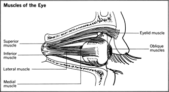
In addition to the tiny muscles within the eye that control the opening of the pupil and the shape of the lens, we also have a number of elegant muscles that control the movements of each eyeball, and make both eyeballs move together in unison.
The movements of each eyeball are affected by six muscles attached to its top, bottom, and sides. The teamwork between these muscles—some contracting while others relax—allows the eye to move from side to side, up and down, and at all intermediate angles (obliquely). One of our eyes is always a dominant or leading eye; that is, its movements are always followed by the other eye.
Our protective eyelids, of course, are controlled by opening and closing muscles that lie outside the eye proper. These muscles can function both voluntarily and involuntarily.
Lubrication and Hygiene of Eye
Without the moisture provided by tears, our eyeball would scrape excruciatingly on the inside lining ( conjunctiva ) of the eyelid. In addition to lubrication, tears also have a cleansing action, not only because they supply water for washing and rinsing but also because they contain a mild germicide called lysozyme that kills bacteria and other potentially harmful microbes.
Tears are produced by the lacrimal glands above the eyeball, just under the eyebrow, a bit further toward the temple side than the nose side. They are discharged from several short ducts and spread over the surface of the eyeball by blinking. The conjunctival sac at the bottom inner (nose) side of the eye—visible in the mirror as a pinkish flap of tissue—serves as a collecting pool for tears; from there, they drain down a duct into the nasal cavity. This is why somebody who is crying also snuffles and must blow his nose.
There is another tiny drainage network in the eye, located at the interconnection of the cornea and iris, which serves to keep the fluid pressure of the space filled by the aqueous humor within normal limits. Drainage of this area is through microscopic conduits called the canals of Schlemm . Improper drainage can cause build-up of pressure, such as occurs in glaucoma, and impairment or loss of vision.