
The Special Senses - Design: parts of the special senses
The abilities to see, to hear, to smell, and to taste are all possible because of sensory receptors, or special nerve cells or endings of the peripheral nervous system (part of the nervous system consisting mainly of nerves that extend from the brain and spinal cord to areas in the rest of the body). Sensory receptors respond to a stimulus by converting that stimulus into a nerve impulse. The impulse is then carried by sensory nerves to a specific part of the brain, where the sensation of sight, sound, smell, or taste is perceived or "felt."
Sensory receptors are classified according to the type of stimulus that arouses or excites them. The receptors for the sense of sight are photoreceptors that are sensitive to light. The receptors for the sense of hearing are mechanoreceptors that are sensitive to sound waves or vibrations. The receptors for the senses of smell and taste are chemoreceptors that are sensitive to various chemicals.
The special sensory receptors for sight and hearing are located in large, complex sensory organs—the eyes and the ears. Those for smell and taste are located in organs that function in other systems—the nose in the respiratory system and the mouth in the digestive system.
Anatomy of the eye
The eye is the organ of sight or vision. Each eye works with the brain to transform light waves into visual images. Eighty percent of all information received by the human brain comes from the eyes.
- Accommodation (ah-kah-mah-DAY-shun):
- Process of changing the shape of the lens of the eye to keep an image focused on the retina.
- Aqueous humor (AYE-kwee-us HYOO-mer):
- Tissue fluid filling the cavity of the eye between the cornea and the lens.
- Binocular vision (by-NOK-yoo-lur VI-zhun):
- Ability of the brain to create one image from the slightly different images received from each eye.
- Ceruminous glands (suh-ROO-mi-nus GLANDZ):
- Exocrine glands in the skin of the auditory canal of the ear that secrete earwax or cerumen.
- Chemoreceptors (kee-moe-re-SEP-terz):
- Receptors sensitive to various chemicals substances.
- Choroid (KOR-oid):
- Middle, pigmented layer of the eye.
- Ciliary body (SIL-ee-air-ee BAH-dee):
- Circular muscle that surrounds the edge of the lens of the eye and changes the shape of the lens.
- Cochlea (KOK-lee-ah):
- Spiral-shaped cavity in the inner ear that contains the receptors for hearing in the organ of Corti.
- Cones:
- Photoreceptors in the retina of the eye that detect colors.
- Cornea (KOR-nee-ah):
- Transparent front portion of the sclera of the eye.
- Conjunctiva (kon-junk-TIE-vah):
- Mucous membrane lining the eyelids and covering the front surface of the eyeball.
- Eardrum (EER-drum):
- Thin membrane at the end of the outer ear that vibrates when sound waves strike it.
- Eustachian tube (yoo-STAY-she-an TOOB):
- Slender air passage between the middle ear cavity and the pharynx, which equalizes air pressure on the two sides of the eardrum.
- External auditory canal (ex-TER-nal AW-di-tor-ee ka-NAL):
- Also called the ear canal, the tunnel in the ear between the pinna and eardrum.
- Gustation (gus-TAY-shun):
- The sense of taste.
- Gustatory cells (GUS-ta-tor-ee CELLS):
- Chemoreceptors located within taste buds.
- Iris (EYE-ris):
- Pigmented (colored) part of the eye between the cornea and lens made of two sets of smooth muscle fibers.
- Lacrimal gland (LAK-ri-muhl GLAND):
- Gland located at the upper, outer corner of each eyeball that secretes tears.
- Lens:
- Clear, oval, flexible structure behind the pupil in the eye that changes shape for the focusing of light rays.
- Mechanoreceptors (mek-ah-no-re-SEP-terz):
- Receptors sensitive to mechanical or physical pressures such as sound and touch.
- Olfaction (ol-FAK-shun):
- The sense of smell.
- Olfactory epithelium (ol-FAK-ter-ee ep-e-THEE-leeum):
- Section of mucous membrane in the roof of the nasal cavity that contains odor-sensitive olfactory nerve cells.
- Organ of Corti (OR-gan of KOR-tee):
- Structure in the cochlea of the inner ear that contains the receptors for hearing.
- Ossicles (OS-si-kuls):
- Three bones of the middle ear: hammer, anvil, and stirrup.
- Papillae (pah-PILL-ee):
- Projections on the tongue that contain taste buds.
- Photoreceptors (fo-to-re-SEP-terz):
- Receptors sensitive to light.
- Pinna (PIN-nah):
- Commonly referred to as the ear, the outer, flaplike portion of the ear.
- Pupil (PYOO-pil):
- Opening in the center of the iris though which light passes.
- Receptors (re-SEP-terz):
- Specialized peripheral nerve endings or nerve cells that respond to a particular stimulus such as light, sound, heat, touch, or pressure.
- Retina (RET-i-nah):
- Innermost layer of the eyeball that contains the photoreceptors—the rods and cones.
- Rods:
- Photoreceptors in the retina of the eye that detect the presence of light.
- Saccule (SAC-yool):
- Membranous sac in the vestibule of the inner ear that contains receptors for the sense of balance.
- Sclera (SKLER-ah):
- Outermost layer of the eyeball, made of connective tissue.
- Semicircular canals (sem-eye-SIR-cue-lar ka-NALZ):
- Three oval canals in the inner ear that help to maintain balance.
- Taste buds:
- Structures on the papillae of the tongue that contain chemoreceptors that respond to chemicals dissolved in saliva.
- Utricle (YOO-tri-kuhl):
- Membranous sac in the vestibule of the inner ear that contains receptors for the sense of balance.
- Vestibule (VES-ti-byool):
- Bony chamber of the inner ear that contains the utricle and the saccule.
- Vitreous humor (VIT-ree-us HYOO-mer):
- Transparent, gellike substance that fills the cavity of the eye behind the lens.
The human eyeball is about 0.9 inch (2.3 centimeters) in diameter and is not perfectly round, being slightly flattened in the front and back. Only about one-sixth of an eye's front surface can normally be seen. The rest of the eye is enclosed and protected by a cushion of fat and the walls of the orbit, a cavity in the skull formed by facial and cranial bones. The eye wall consists of three covering layers: the sclera, the choroid, and the retina.
THE SCLERA. The sclera, the outer layer made of fibrous connective tissue, encases and protects the eyeball. The visible portion of the sclera is seen as the "white" of the eye. When that portion is irritated, the small blood vessels contained in the layer enlarge, producing a "bloodshot eye." In the center of the visible portion of the sclera is the cornea, which projects slightly forward. The cornea is transparent and has no capillaries. It is the "window" or the first part of the eye through which light enters. A delicate mucous membrane, the conjunctiva, covers the cornea and visible portion of the sclera. It secretes mucus to lubricate the eyeball and keep it moist.
THE CHOROID. The choroid is a thin membrane lying underneath the sclera. It is composed of a dark pigment that absorbs light within the eye (preventing glare) and numerous blood vessels that nourish the internal tissues of the eye. At the front end of the choroid is the ciliary body. Running like a ring around the visible portion of the eye, the ciliary body connects the choroid with the iris. The ciliary body contains muscles that are connected by ligaments to the lens behind the iris.
The iris is the visible portion of the choroid. It gives the eye its color, which varies depending on the amount of pigment present in the iris. Dense pigment makes the iris brown, while little pigment makes the iris blue. If there is no pigment the iris is pink, as in the eye of a white rabbit. The rounded opening in the center of the iris is the pupil, through which light passes. In bright light, muscles in the iris constrict the pupil, reducing the
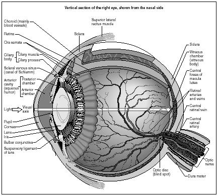
amount of light entering the eye. Conversely, the pupil dilates (enlarges) in dim light, increasing the amount of light entering. Extreme fear, head injuries, and certain drugs can also dilate the pupil.
THE LENS. The lens is a crystal-clear, oval, flexible body that is biconvex (curving outward on both surfaces). It is made up of approximately 35 percent protein and 65 percent water. The entire surface of the lens is smooth and shiny, contains no blood vessels, and is encased in an elastic membrane. The lens sits behind the iris and focuses light on the retina. In addition to holding the lens in place, the muscles of the ciliary body contract and relax, causing the lens to either fatten or become thin. As the shape of the lens changes, so does its focus.
THE RETINA. The retina, the innermost layer of the eye, is thin, delicate, sensory tissue composed of layers of light-sensitive nerve cells. The retina begins at the ciliary body (not at the front of the eye) and encircles the entire interior portion of the eye. Rods and cones are the photoreceptors of the retina. In each eye there are about 126 million rods and 6 million cones.
Rods function chiefly in dim light, allowing limited night vision: it is with rods that a person sees the stars. Rods cannot detect color (that is why objects in dim light appear in shades of gray), but they are the first cells to detect movement. They are most abundant toward the edge of the retina and provide people with peripheral (or side) vision. Cones function best in bright light and are sensitive to color. They are most abundant in the center of the retina. Scientists believe three types of cones—red, blue, and green—exist in the eye. The perception of different colors is the result of the stimulation of various combinations of these three types.
CAVITIES AND FLUIDS OF THE EYE. Between the cornea and the lens is a small cavity. This cavity is filled with a clear watery fluid known as aqueous humor, formed by capillaries in the ciliary body. This fluid aids good vision by helping maintain eye shape, providing support for the internal structures, supplying nutrients to the lens and cornea, and disposing of cellular wastes produced by the eye.
The large cavity in back of the lens (the center of the eyeball) is filled with a transparent, gellike substance called vitreous humor. Light passing through the lens on its way to the retina passes through the vitreous humor. The vitreous humor is 99 percent water and contains no cells. It helps to maintain the shape of the eye and support its internal components.
The amount of pigment in the iris is what determines its color. In newborns, most of the pigment is concentrated in the folds of the iris. Since only a little bit of pigment exists on the visible portion of the iris, it appears blue. When a baby is a few months old, the rest of the pigment begins moving to the surface of the iris, giving the baby his or her permanent eye color.
ACCESSORY STRUCTURES OF THE EYE. The lacrimal gland, which lies immediately above each eyeball at the outer corner of the eye socket, produces tears. Tears are mostly water, but also contain antibodies and an enzyme that prevents the growth of most bacteria on the wet, warm surface of the eye. Tears flow through numerous ducts from the lacrimal gland to the area beneath the upper eyelid. Blinking spreads the tears across the cornea's outside surface, keeping it moist and clean. Tear fluid then either evaporates or drains into two small pores in the inner corner of the eye that connect to a larger duct, which eventually drains tears into the nasal cavity.
Eyelids and eyelashes help to protect the eye. The blinking movement of eyelids keeps the front surface of the eye lubricated and free from dust and dirt. The rate of blinking varies. On average, the eye blinks once every five seconds (or 17,280 times a day or 6.3 million times a year). The eyelids can also close firmly to protect the eye. Eyelashes, hairs that project from the border of each eyelid, help to keep dust, dirt, and insects out of the eye.
Extending from the bony surface of the orbit to the outside of the eyeball are six small muscles that contract and relax, allowing the eye to move in various directions. Four of the muscles move the eyeball up and down and side to side. The other two muscles rotate the eye.
Anatomy of the ear
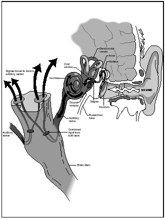
The human ear is the organ responsible for hearing and for equilibrium or balance. The ear consists of three regions or areas: the outer (external) ear, the middle ear, and inner (internal) ear. The mechanoreceptors for hearing and balance are all found in the inner ear.
THE OUTER EAR. The outer ear collects external sounds and funnels them through the auditory system to the eardrum. The outer ear is composed of three parts—the pinna (or auricle), the external auditory canal, and the eardrum (tympanic membrane).
What are commonly called ears—the two flaplike structures on either side of the head—are actually the pinnas of the outer ear. Pinnas are skin-covered cartilage, not bone, and are therefore flexible. In many species of animals, the pinnas act to collect and funnel sound waves into the external auditory canal. In humans, however, the pinnas do not serve this purpose. In fact, humans could lose their pinnas and hearing would not be adversely affected.
Floaters are semi-transparent or dark little specks that float across the field of vision and can be mistaken for flies in the room. Some floaters originate with red blood cells that have leaked out of the retina. The blood cells swell into spheres—some forming strings—and float around the areas of the retina. Others are shadows cast by microscopic structures in the vitreous humor.
A sudden appearance of dark floaters, if accompanied by bright little flashes, could indicate that the retina has detached, a serious problem that requires medical treatment.
The external auditory canal or ear canal is a passageway that begins at the pinna and extends inward and slightly upwards, ending at the
eardrum. In the adult human it is lined with skin and hairs and is approximately 1 inch (2.5 centimeters) long. The outer one-third of the canal is lined with wax-producing ceruminous glands and fine hairs. The purpose of the earwax and hairs is to protect the eardrum by trapping dirt and foreign bodies and keeping the canal moist.
The eardrum or tympanic membrane is a thin, concave membrane stretched across the inner end of the auditory canal much like the skin covering the top of a drum. The eardrum marks the border between the outer ear and middle ear. In the adult human, the eardrum has a total area of approximately 0.1 square inch (0.6 square centimeter). The middle point of the eardrum—called the umbo—is attached to the stirrup, the first of three bones contained within the middle ear.
THE MIDDLE EAR. The middle ear transmits sound from the outer ear to the inner ear. The middle ear consists of an oval, air-filled space approximately 0.1 cubic inch (2 cubic centimeters) in volume. Contained in this space are three tiny bones called ossicles. Because of their shapes, the three ossicles are known as the hammer (malleus), the anvil (incus), and the stirrup (stapes).
Connecting the middle ear to the throat is the eustachian tube. This tube is normally closed, opening only as a result of muscle movement during yawning, sneezing, or swallowing. The eustachian tube causes air pressure in the middle ear to match the air pressure in the outer ear.
Hearing aids are tools that amplify sound for people who have a hard time hearing. Millions of hearing aids are sold annually, especially to people over the age of sixty-five. More than 1,000 different models are available in the United States.
A typical hearing aid contains a microphone that picks up sounds and changes them into electric signals. The hearing aid's amplifier increases the strength of the electric signals. Then the receiver converts the signals back into sound waves that can be heard by the wearer.
The entire mechanism is housed in an ear mold that fits snugly in the ear canal. The power to run the electronic parts is provided by a small battery. There are a variety of designs to fit the needs of the wearer, some small enough to be completely concealed by the ear canal.
Many modern hearing aids have miniature computer chips that allow the aid to selectively boost certain frequencies. This means that a person could wear such a hearing aid to a loud party and screen out unwanted background noise while tuning in to a private conversation. The hearing aid can also be programmed to conform to a person's specific hearing loss. Some models can be further programmed to allow the wearer to choose different settings depending on the noise of the surroundings.
The most noticeable example of eustachian tube function occurs when there is a quick change in altitude, such as when a plane takes off. Prior to takeoff, the pressure in the outer ear is equal to the pressure in the middle ear. When the plane gains altitude, the air pressure in the outer ear decreases, while the pressure in the middle ear remains the same, causing the ear to feel "plugged." In response to this the ear may "pop." The popping sensation is actually the quick opening and closing of the eustachian tube and the equalization of pressure between the outer and middle ear.
THE INNER EAR. The inner ear is responsible for interpreting and transmitting sound and balance sensations to the brain. The inner ear, located just behind the eye socket, is small (about the size of a pea) and complex in shape. With its series of winding interconnected chambers, it has been called a labyrinth. The main components of the inner ear are the vestibule, semicircular canals, and the cochlea.
The vestibule, a round open space, is the central structure within the inner ear. The vestibule contains two membranous sacs—the utricle and the saccule (the saccule is the smaller of the two). These sacs, lined with tiny hairs and attached to nerve fibers, function as an individual's chief organs of balance.
Attached to the vestibule are three loop-shaped, fluid-filled tubes called the semicircular canals. These canals, arranged perpendicular to each other, are a key part of the vestibular system. Two of the canals help the body maintain balance when it is moving vertically, such as in falling and jumping. The third maintains horizontal balance, as when the head or body rotates.
The cochlea is the organ of hearing. The cochlea consists of a bony, snail-like shell that contains three separate fluid-filled ducts or canals. The middle canal contains the basilar membrane, which holds or supports the organ of Corti, named after Italian anatomist Alfonso Giacomo Gaspare Corti (1822–1876) who discovered it. The organ contains over 20,000 hair cells (mechanoreceptors) connected at their base to the auditory nerve. The organ is the site where sound waves are converted into nerve impulses, which are then sent to the brain along the auditory nerve.
Anatomy of the sense of smell
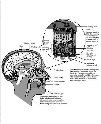
Smell, called olfaction, is the ability of an organism to sense and identify a substance by detecting tiny amounts of the substance that evaporate and produce an odor. Smell is the most important sense for most organisms.
In humans, the sense of smell differs from the other senses (sight, hearing, and taste) in its directness. People actually smell microscopic bits or chemicals of a substance that have evaporated and made their way through the nostrils into the nasal cavity. In the roof of the nasal cavity is a section of mucous membrane called the olfactory epithelium. It covers an area of roughly 0.75 square inch (4.8 square centimeters), or about the size of a postage stamp.
The olfactory epithelium contains millions of odor-sensitive olfactory receptor cells (chemoreceptors) that are connected to the olfactory nerves. The olfactory receptors have long olfactory hairs that protrude outward from the epithelium. Beneath the olfactory epithelium lie olfactory glands that produce mucus that covers the epithelium and bathes the olfactory hairs. The mucus keeps the area moist and clean and prevents the buildup of potentially harmful or overpowering chemicals.
Anatomy of the sense of taste
Taste, called gustation, is the sense for determining the flavor of food and other substances (taste comes from the Latin word taxare , meaning "to touch" or "to feel"). It is one of the two chemical senses (the other being smell) and it is stimulated when taste buds on the tongue come in contact with certain chemicals. The sense of taste is also influenced by the smell and texture of substances, hereditary factors, culture, and familiarity with specific taste sensations.
Normally, when people experience the world through their senses, they do so in an orderly fashion. They see with their eyes, hear with their ears, and taste and smell with the chemoreceptors in their mouths and noses.
For some people, however, the basic rules of sensory perception do not apply. They tend to perceive stimuli not only with the sense for which it was intended, but with others as well—sight mingles with sound, taste with touch. They may see musical notes as color hues or feel flavors as different textures on the skin.
This rare condition is known as synesthesia (sines-THE-zee-ah), and the people who have it as synesthetes. Women are about six times as likely as men to be synesthetes.
Some scientists believe that the condition is the result of associations learned at an early age. Other scientists disagree, believing that a unique physical condition exists in the brains of synesthetes. Some brain studies have shown that during synesthetic experiences, blood flow to some parts of the brain decreases. Normally, that blood flow would have been increased by sensory stimuli. Synesthesia also appears to run in families, leading some scientists to theorize that it has a genetic basis.
Clusters of small organs called taste buds are located in the mouth. Of the almost 10,000 taste buds, most are located on the upper surface of the tongue (a few are located on the soft palate and on the inner surface of the cheeks). Taste buds (named so because under the microscope they look similar to plant buds) lie in small projections on the tongue called papillae. Within the taste buds are taste receptors known as gustatory cells. Each gustatory cell projects slender taste hairs into the surrounding fluids through a narrow taste pore. As food is broken down in the mouth, these receptors come into contact with chemicals dissolved in the saliva. They then send messages along nerves to the brain, which interprets the flavor as sweet, sour, salty, or bitter.
Taste buds for all four taste groups can be found throughout the mouth, but specific kinds of buds are concentrated in certain areas (the areas tend to overlap each other). Sweetness is detected by taste buds on the tip of the tongue. The buds for sour tastes are on the sides of the tongue. Those for salty are toward the front. Bitter taste buds are located on the back of the tongue. Bitterness can make many people gag, which is a defense mechanism. Since many natural poisons and spoiled foods are bitter, gagging helps prevent poisoning.
New taste buds are produced every three to ten days to replace the ones worn out by scalding or frozen foods. As people grow older, their taste buds are replaced at a slower rate, and more of a substance is needed to experience its full flavor.
Comment about this article, ask questions, or add new information about this topic: