
The Respiratory System - Design: parts of the respiratory system
Breathing describes the process of inhaling and exhaling air. The exchange of gases (oxygen and carbon dioxide) between living cells and the environment is a process known as respiration. The respiratory system, which controls breathing and respiration, consists of the respiratory tract and the lungs.
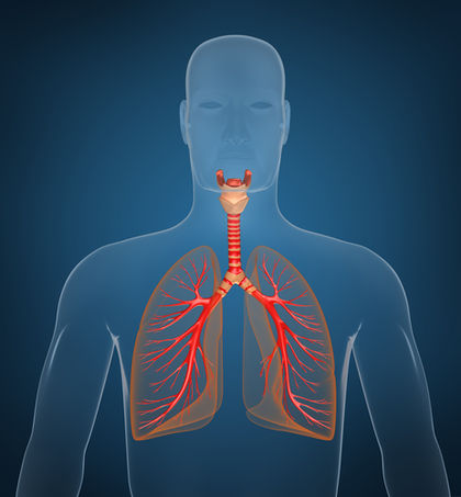
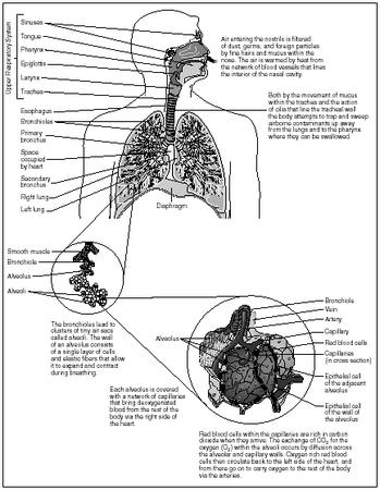
The respiratory tract cleans, warms, and moistens air on its way to the lungs. The tract can be divided into an upper and a lower part. The upper part consists of the nose, nasal cavity, pharynx (throat), larynx, and upper part of the trachea (windpipe). The lower part consists of the lower part of the trachea, bronchi, and lungs (which contain bronchioles and alveoli).
The nose and nasal cavity
The nose is the only external part of the respiratory system. It is made of bone and cartilage (tough connective tissue) and is covered with skin. The two openings to the outside, called nostrils, allow air to enter or leave the body during breathing. The nostrils are lined with coarse hairs that prevent large particles such as dust, insects, and sand from entering.
The nostrils open into a large cavity, the nasal cavity. This cavity is divided into right and left cavities by a thin plate of bone and cartilage called the nasal septum. The hard portion of the palate forms the floor of the entire nasal cavity, separating it from the mouth or oral cavity below. Three flat, spongy folds or plates project toward the nasal septum from the sides of the nasal cavity. These plates, called nasal conchae, help to slow down the passage of air, causing it to swirl in the nasal cavity.
- Alveoli (al-VEE-oh-lie):
- Air sacs of the lungs.
- Breathing (BREETH-ing):
- Process of inhaling and exhaling air.
- Bronchi (BRONG-kie):
- Largest branch of the bronchial tree between the trachea and bronchioles.
- Bronchial tree (BRONG-key-uhl TREE):
- Entire system of air passageways within the lungs formed by the branching of bronchial tubes.
- Bronchioles (BRONG-key-ohls):
- Smallest of the air passageways within the lungs.
- Epiglottis (ep-i-GLAH-tis):
- Flaplike piece of tissue at the top of the larynx that covers its opening when swallowing is occurring.
- Esophagus (i-SOF-ah-gus):
- Muscular tube connecting the pharynx and stomach.
- Exhalation (ex-ha-LAY-shun):
- Also known as expiration, the movement of air out of the lungs.
- Glottis (GLAH-tis):
- Opening of the larynx between the vocal cords.
- Hemoglobin (HEE-muh-glow-bin):
- Iron-containing protein pigment in red blood cells that can combine with oxygen and carbon dioxide.
- Inhalation (in-ha-LAY-shun):
- Also known as inspiration, the movement of air into the lungs.
- Larynx (LAR-ingks):
- Organ between the pharynx and trachea that contains the vocal cords.
- Lungs:
- Paired breathing organs.
- Nasal cavity (NAY-zul KAV-i-tee):
- Air cavity in the skull through which air passes from the nostrils to the upper part of the pharynx.
- Nasal conchae (NAY-zul KAHN-kee):
- Flat, spongy plates that project toward the nasal septum from the sides of the nasal cavity.
- Nasal septum (NAY-zul SEP-tum):
- Vertical plate made of bone and cartilage that divides the nasal cavity.
- Nose:
- Part of the human face that contains the nostrils and organs of smell and forms the beginning of the respiratory tract.
- Nostril (NOS-tril):
- Either of the two external openings of the nose.
- Paranasal sinuses (pair-a-NAY-sal SIGH-nus-ez):
- Air-filled chambers in the bones of the skull that open into the nasal cavity.
- Pharynx (FAR-inks):
- Short, muscular tube extending from the mouth and nasal cavities to the trachea and esophagus.
- Pleura (PLOOR-ah):
- Membrane sac covering and protecting each lung.
- Pulmonary surfactant (PULL-mo-nair-ee sir-FAK-tent):
- Oily substance secreted by the alveoli to prevent their walls from sticking together.
- Respiration (res-pe-RAY-shun):
- Exchange of gases (oxygen and carbon dioxide) between living cells and the environment.
- Trachea (TRAY-key-ah):
- Also known as the windpipe, the respiratory tube extending from the larynx to the bronchi. The nasal cavity is lined by mucous membrane containing microscopic hairlike structures called cilia. The cells of the membrane produce mucus, a
thick, gooey liquid. As the nasal conchae cause air to swirl in the nasal cavity, the mucus moistens the air and traps any bacteria or particles of air pollution. The cilia wave back and forth in rhythmic movement, and pieces of mucus with their trapped particles are swept along to the throat. The mucus is then either spat out or (more often) swallowed. Any bacteria present in the swallowed mucus is destroyed by the hydrochloric acid in the gastric juice of the stomach.
Air is not only moistened in the nasal cavity but warmed, as well. A rich network of thin-walled capillaries permeates the mucus membrane (especially the uppermost concha), and the incoming air is warmed as it passes over the vessels. When air finally reaches the lungs, it is similar to the warm, damp air found in the tropics.
The bones that surround the nasal cavity contain hollow spaces known as paranasal sinuses. The sinuses are also lined with mucous membrane containing cilia. The mucus produced in the sinuses drains into the nasal cavity. The main functions of the sinuses are to lighten the skull and to provide resonance (sound quality) for the voice.
The pharynx
The pharynx or throat is a short, muscular tube extending about 5 inches (12.7 centimeters) from the nasal cavity and mouth to the esophagus and trachea. It serves two separate systems: the digestive system (by allowing the passage of solid food and liquids) and the respiratory system (by allowing the passage of air).
The larynx
The larynx, commonly called the voice box, forms the upper part of the trachea. The larynx is made of nine pieces of cartilage connected by ligaments. The largest of these cartilages is the shield-shaped thyroid cartilage, which may protrude at the front of the neck, forming the so-called Adam's apple. The upper cartilage is the epiglottis, a flaplike piece of tissue. During swallowing, the larynx rises up and the epiglottis folds down to cover the glottis, or the larynx's opening. This prevents food or liquids from passing into the lower respiratory tract.
Mucous membrane lines the larynx. A pair of elastic folds in that lining form the vocal cords. During silent breathing, the vocal cords lie against the walls of the larynx. During speech, the cords are stretched across the opening of the larynx and air that passes through causes them to vibrate, generating sound waves. Various muscles produce tension on the cords, making them tighter (shorter) or looser (longer). The tighter the tension, the higher the pitch of the sound produced. Since men's larynges tend to be larger than women's, their vocal cords tend to be thicker and longer. The male voice thus tends to be lower in pitch.
The trachea
The trachea is a tough, flexible tube about 1 inch (2.5 centimeters) in diameter and 4.5 inches (11.4 centimeters) in length. Located in front of the esophagus, it is the principal tube that carries air to and from the lungs. The walls of the trachea are supported by 16 to 20 C-shaped cartilage rings. Elastic fibers in the tracheal walls allow the trachea to expand and contract during breathing, while the cartilage rings prevent it from collapsing. Mucous membrane containing cilia lines the trachea. The mucus produced by the membrane traps dust particles and other debris. The cilia move continuously in a direction opposite that of the incoming air, helping propel the mucus away from the lungs to the throat where it can be swallowed or spat out.
The bronchi
The trachea divides behind the sternum (breastbone) to form a right and left branch called primary bronchi (singular: bronchus). Each bronchus passes into a lung—the right bronchus into the right lung and the left bronchus into the left lung. The right bronchus is wider, shorter, and straighter than the left. As a result, accidentally inhaled objects (such as pieces of food) most often enter the right primary bronchus.
By the time incoming air reaches the primary bronchi, it is warm, moistened, and cleansed of most particles or other impurities.
The lungs
The lungs are two broad, cone-shaped organs located on either side of the heart in the thoracic or chest cavity. They extend from the collarbones to the diaphragm, a membrane of muscle separating the thoracic cavity from the abdominal cavity. The base of each lung rests directly on the diaphragm. The rib cage forms a wall around the lungs, protecting them.
At birth, the lungs are pale pink in color. As people age, their lungs grow darker. The inhaling of dirt and other particles increases this aging process, even scarring the delicate tissue of the lungs.
Each lung is divided into lobes separated by deep grooves or fissures. The right lung, which is larger, is divided into three lobes. The left lung is divided into only two lobes. Combined, the two soft and spongy lungs weigh about 2.5 pounds (1.1 kilograms).
A membrane sac, called the pleura, surrounds and protects each lung. One layer of the pleura attaches to the wall of the thoracic cavity; the other layer encloses the lung. A fluid (pleural fluid) between the two membrane layers reduces friction and allows smooth movement of a lung during breathing.
After the bronchi enter the lungs, they subdivide repeatedly into smaller and smaller bronchi or branches. Eventually they form thousands of tiny branches called bronchioles, which have a diameter of about 0.02 inch (0.5 millimeter). This branching network of bronchial tubes within the lungs is called the bronchial tree.
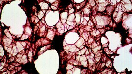
The bronchioles branch to form even smaller passageways that open into clusters of cup-shaped air sacs called alveoli (singular: alveolus). The average person has a total of about 700 million alveoli (which resemble clusters of grapes) in his or her lungs. These provide an enormous surface area-roughly the size of a tennis court—for gas exchange. A network of capillaries surrounds each alveolus. As blood passes through these vessels and air fills the alveoli, the exchange of gases takes place: oxygen passes from the alveoli into the capillaries while carbon dioxide passes from the capillaries into the alveoli.
The membranes of the alveoli are extremely delicate and thin to allow the gases to pass easily through them. The inner lining of those membranes is coated with a thin layer of tissue fluid (a gas must be dissolved in a liquid in order to enter or leave a cell). To prevent the walls of the alveoli from sticking together (like the inside walls of a wet plastic bag), cells in the alveoli also produce an oily secretion, called pulmonary surfactant, that mixes with the tissue fluid (pulmonary refers to anything relating to or affecting the lungs).
Comment about this article, ask questions, or add new information about this topic: