The Nervous System - Workings: how the nervous system functions

Reading a book, walking through a field, playing a musical instrument, digesting a holiday meal, remembering a lost relative—the nervous system regulates all of the body's activities, from the simplest to the most complex. In order to perceive and to respond to the world around us and the changes within us, the body's tissues, organs, and organ systems must function. In order for those body parts to function, they must be stimulated and regulated by nerve impulses.
Neurons have the ability to respond to a stimulus and convert it into a nerve impulse. They also have the ability to transmit that impulse to other neurons or the cells of muscles or glands.
Transmission of nerve impulses
In neurons, information travels in the form of nerve impulses that are conducted along axons. In myelinated axons, a nerve impulse can travel up to about 325 feet (100 meters) per second. In unmyelinated axons, the impulse travels much slower, about 1.5 feet (0.5 meter) per second.
Impulses do not travel in and between neurons like electric currents through telephone wires. For nerve impulses to be transmitted throughout the body, electrochemical reactions must occur in neurons. As stated earlier, dendrites are the points through which signals or impulses from adjacent neurons enter a particular neuron. If a dendrite of a neuron is stimulated, electrical and chemical changes take place throughout the cell.
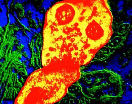
Every neuron communicates with other neurons or with other types of cells. Neurons that transmit impulses to other neurons do not actually touch one another. The small space or gap where the impulse passes between the axon of one neuron and a dendrite of the next neuron is known as the synapse. A synapse measures about 0.000001 inch (0.0000025 centimeter).
When a neuron is inactive or resting, the tissue fluid that surrounds it contains more positive ions than are inside the neuron (an ion is an atom or group of atoms that has an electrical charge, either positive or negative). The major positive ions outside the cell are sodium; the major positive ions inside the cell are potassium. Because there are more positive ions outside the neuron, its internal surface is slightly negative. As long as the outside remains positive and the inside negative, the neuron remains at rest.
When a dendrite of a neuron is stimulated, tiny "gates" in the membrane of the dendrite and cell body begin to open and close. According to the laws of diffusion, molecules always move from an area where they exist in greater numbers to an area where they exist in lesser numbers. So, when these gates are open, sodium ions flow into the cell body. This temporary movement of ions changes the electrical charges on the membrane of the cell—positive on the inside, negative on the outside. This results in an electrical charge or nerve impulse on the surface of the cell. The impulse rapidly passes from the dendrite, down the length of the cell body, and along the length of the axon. An all-or-nothing response, the nerve impulse never goes partway along a neuron, but along its entire length.
The electrical charges (nerves impulses) created by the billions of neurons in the brain combine to generate an electrical field. That field can be measured by a machine called an electroencephalograph. The machine records the electrical activity as horizontal zigzag patterns on a screen or a page. Those patterns are known as brain waves.
As different regions of the brain are stimulated or are quieted down, the brain wave patterns change. There are four major types of brain waves. Alpha waves occur in normal, healthy adults who are awake but relaxed. The waves produce regular, fast patterns, about 8 to 13 cycles per second. Beta waves result when a person is concentrating or thinking about something. They produce patterns that are small and very fast, about 13 to 30 cycles per second. Theta waves are found in the brains of children and in adults who are under emotional stress. In adults, theta waves may also indicate a brain disorder. The patterns of theta waves are large and slow, about 4 to 8 cycles per second. The largest and slowest-moving waves are delta waves. These regular-patterned waves appear in the brains of infants and in the brains of sleeping adults. They are also found in the brains of individuals who have suffered brain damage.
By comparing the brain waves of a person to those found in normal, healthy individuals, physicians can determine if the brain of that person has been injured or infected by a disease.
As soon as the gates on the membrane of the neuron open, they close and the body restores the correct balance of sodium and potassium ions inside and outside the neuron. Only after the balance has been restored and the neuron is at rest can another impulse be conducted along the neuron.
NEUROTRANSMITTERS. When the impulse or electrical current has reached the terminal branches or end of the axon, it stimulates the branches to release chemicals known as neurotransmitters. As their name suggests, neurotransmitters are the mechanisms by which a nerve impulse travels from one neuron to another or to body cells.
Once released from an axon, a neurotransmitter drifts across the synapse to a second neuron. When it has reached that cell, it attaches itself to specialized parts, called receptors, in the dendrites of the second neuron. This act of attaching creates the stimulus for dendrites in the second neuron. Those

dendrites then respond to this stimulus just as the first neuron responded to its stimulus, and the nerve impulse continues.
Scientists have identified a number of neurotransmitters, including dopamine, serotonin, and acetylcholine. Each neurotransmitter occurs in certain types of neurons and has specific functions. It can either start an action or stop it, such as causing a muscle to contract or a gland to stop secreting. For example, acetylcholine is the neurotransmitter released at the terminal branches of motor neurons that come in close contact with muscle fibers (in this case, the synapse is called the neuromuscular junction). When acetylcholine attaches to receptors on the membrane of the muscle fiber, it triggers an electrical charge that quickly travels from one end of the muscle fiber to the other, causing it to contract.
The transmission of a nerve impulse—first along a neuron, then to another neuron—is an electrochemical event. The impulse traveling along a neuron is electrical, but the transfer of that impulse (the stimulation of the next neuron by neurotransmitters) is chemical. The actions involved in the creation and transfer of a nerve impulse ensure that it can travel in one direction only in a neuron—from dendrite through axon.
Even though nerve impulses travel in one direction only, a neuron can have as many as 100,000 synapses connecting it to other neurons. Thus, a neuron can receive and transmit many impulses over a variety of pathways, connecting with many different neurons at the same time. The path an impulse takes, from a particular neuron to another one, determines the meaning of that impulse and the action it evokes.
Reflexes
It is not always necessary for a nerve impulse traveling along sensory nerves to reach the brain before a response causes the body to react in some way. When a stimulus causes a response that is involuntary, rapid, and predictable, that response is known as a reflex. Reflexes are classified according to the systems of the PNS: autonomic reflexes and somatic reflexes. Autonomic reflexes control the activity of the smooth muscles, heart, and glands. They also regulate complex body functions such as digestion, blood pressure, sweating, and swallowing. Somatic reflexes control skeletal muscles. In general, reflexes control much of what the body must do every day.
The pathway a nerve impulse travels when a reflex is initiated is called the reflex arc. A typical reflex action begins when a sensory receptor (in the skin, sense organ, or other internal organ) is activated by some kind of stimulus. The receptor generates an impulse in a sensory neuron and the impulse then travels along other sensory neurons to the spinal cord. In the gray matter of the spinal cord, the impulse passes from a sensory neuron through an interneuron into a motor neuron. The motor neuron then transmits the impulse through other motor neurons to a muscle or gland, where some type of response occurs.
For example, if a person touches a hot stove, that person immediately pulls his or her hand away. Heat and pain receptors in the skin were stimulated to send impulses to the spinal cord, where they were transferred to motor neurons that eventually connected to muscles in the hand, which were stimulated to contract and pull the hand away. Although it may seem that the pain caused by the hot stove led to the quick withdrawal of the hand, the movement actually occurred milliseconds before the brain became aware of the pain and initiated some response. This is an example of a somatic reflex.
Reflexes such as this one are important in safeguarding the body against potentially harmful changes outside or inside the body. Reflexes are also a good tool in evaluating the condition of the nervous system. If a reflex is exaggerated or even absent, a nervous system disorder may be present. That is why doctors often perform the knee-jerk reflex test during a routine physical examination: a sharp rap on the tendon below the knee should cause the quadriceps muscle on the front part of the thigh to contract, forcing the lower leg to kick outward.
Autonomic nervous system
Much of what occurs in the body every day occurs without an individual being consciously aware. The heart beats, the lungs expand, blood vessels contract and dilate (widen), and the stomach and intestines break down food and move it through the system. These actions and all others that take place without willful control are regulated by the autonomic nervous system (ANS), a part of the peripheral nervous system.
All body systems contribute to homeostasis or the ability of the body to maintain the internal balance of its functions. The minute-to-minute stability of the body, however, is largely dependent on the workings of the ANS.
The ANS is broken down into two subdivisions. The part that keeps body systems running smoothly on a daily basis is called the parasympathetic nervous system. The part that comes into play when emergencies or stressful situations arise is called the sympathetic nervous system. Both subdivisions service the same body organs and use motor nerves to do so (some glands and skin structures receive only sympathetic motor nerves).
The parasympathetic nervous system is in control when the body is at rest. The neurons for this system originate in the brain stem and in the lower region of the spinal cord. The impulses conducted through this system target body organs in an effort to conserve body energy and promote normal digestion and elimination. In the digestive tract, secretions and peristalsis (series of wavelike muscular contractions that move material in one direction through a hollow organ) increase. Heart rate, the force of heart contractions, and blood pressure all decrease. The pupils of the eyes constrict to limit the amount of light entering the body. Kidneys increase their production of urine. Nutrient levels in the blood increase, and cells throughout the body add the extra nutrients to their energy reserves.
The sympathetic nervous system has the opposite effect on the body. It comes into play when an individual is faced with a "fight-or-flight" situation. The neurons for this system originate from the middle of the spinal cord. Their impulses seek to stimulate the body into using its energy. The activity of the digestive and urinary organs decreases. The liver releases glucose (sugar) into the blood for use by the cells as energy. Heart rate and contraction, blood pressure, and blood flow to skeletal muscles all increase. The eyes dilate to let more light in. In short, the sympathetic nervous system prepares the body to respond to some threat, whether that response is to run, to see better, or to think more clearly.

Italian-born American neurobiologist Rita Levi-Montalcini (1909– ) is recognized for her groundbreaking research on nerve cell growth.
She discovered a protein in the human nervous system that she named the nerve growth factor (NGF). For her work, which has proven useful in the study of several disorders (including Alzheimer's disease), Levi-Montalcini received the 1986 Nobel Prize for physiology or medicine (she shared the award with biochemist Stanley Cohen).
After graduating from medical school in Italy in 1936, Levi-Montalcini began to research the nervous system. She conducted experiments on chicken embryos (organisms in their earliest stages of development) in order to study how neurons are differentiated, or how they are formed and assigned a particular function in the developing body. Levi-Montalcini believed that a specific nutrient was essential for neuron growth. In the 1950s, she finally isolated or obtained a sample of the substance that caused neurons to grow and labeled it NGF.
The work that Levi-Montalcini began in the late 1930s has been carried on by researchers who realize the important role that NGF can possibly play in treating degenerative diseases (those in which organs or tissue deteriorate and stop functioning).
The parasympathetic and sympathetic nervous systems rarely work independently of each other. Instead, they often work together, especially in affecting vital organs. Their opposing effects help to maintain the dynamic balance of the internal body.

Comment about this article, ask questions, or add new information about this topic: