The Lymphatic System - Design: parts of the lymphatic system
A network of vessels, tissues, organs, and cells constitute the lymphatic system. Included in this network are lymph vessels, lymph nodes, the spleen, the thymus, and lymphocytes. Running throughout this network is a watery fluid called lymph.
Lymph
Lymph comes from the Latin word lympha , meaning "clear water." Slightly yellowish but clear, lymph is any tissue or interstitial fluid that enters the lymph vessels. It is similar to blood plasma, but contains more white blood cells. Lymph also carries other substances, depending on where it is in the body. In the limbs, lymph is rich in protein, especially albumin. In the bone marrow, spleen, and thymus, lymph contains higher concentrations of white blood cells. And in the intestine, lymph contains fats absorbed during digestion.
Lymph vessels
Lymph vessels, also called lymphatics, carry lymph in only one direction—to the heart. Throughout all the tissues of the body, lymph vessels form a complicated, spidery network of fine tubes. The smallest vessels, called lymph capillaries, have closed or dead ends (unlike vessels in the cardiovascular system, which form a closed circuit). The walls of these capillaries are composed of only a single layer of flattened cells. Material in the interstitial fluid passes easily through the gaps between these cells into the capillaries. Lymph capillaries in the villi (tiny fingerlike projections) of the small intestine are called lacteals. These specialized capillaries transport the fat products of digestion, such as fatty acids and vitamin A.
- Allergen (AL-er-jen):
- Substance that causes an allergy.
- Antibody (AN-ti-bod-ee):
- Specialized substance produced by the body that can provide immunity against a specific antigen.
- Antibody-mediated immunity (AN-ti-bod-ee MEE-deea-ted i-MYOO-ni-tee):
- Immune response involving B cells and their production of antibodies.
- Antigen (AN-ti-jen):
- Any substance that, when introduced to the body, is recognized as foreign and activates an immune response.
- B cell:
- Also called B lymphocyte, a type of lymphocyte that originates from the bone marrow and that changes into antibody-producing plasma cells.
- Cell-mediated immunity (CELL MEE-dee-a-ted i-MYOO-ni-tee):
- Immune response led by T cells that does not involve the production of antibodies.
- Chyle (KILE):
- Thick, whitish liquid consisting of lymph and tiny fat globules absorbed from the small intestine during digestion.
- Edema (i-DEE-mah):
- Condition in which excessive fluid collects in bodily tissue and causes swelling.
- Fever:
- Abnormally high body temperature brought about as a response to infection or severe physical injury.
- Histamine (HISS-ta-mean):
- Chemical compound released by injured cells that causes local blood vessels to enlarge.
- Immunity (i-MYOO-ni-tee):
- Body's ability to defend itself against pathogens or other foreign material.
- Inflammation (in-flah-MAY-shun):
- Response to injury or infection of body tissues, marked by redness, heat, swelling, and pain.
- Interferon (in-ter-FIR-on):
- Protein compound released by cells infected with a virus to prevent that virus from reproducing in nearby normal cells.
- Lacteals (LAK-tee-als):
- Specialized lymph capillaries in the villi of the small intestine.
- Lymph (LIMF):
- Slightly yellowish but clear fluid found within lymph vessels.
- Lymph node:
- Small mass of lymphatic tissue located along the pathway of a lymph vessel that filters out harmful microorganisms.
- Lymphocyte (LIM-foe-site):
- Type of white blood cell produced in lymph nodes, bone marrow, and the spleen that defends the body against infection by producing antibodies.
- Macrophage (MACK-row-fage):
- Large white blood cell that engulfs and destroys bacteria, viruses, and other foreign substances in the lymph.
- Natural killer cell:
- Also known as an NK cell, a type of lymphocyte that patrols the body and destroys foreign or abnormal cells.
- Peyer's patches (PIE-erz):
- Masses of lymphatic tissue located in the villi of the small intestine.
- Phagocyte (FAG-oh-site):
- Type of white blood cell capable of engulfing and digesting particles or cells harmful to the body.
- Phagocytosis (fag-oh-sigh-TOE-sis):
- Process by which a phagocyte engulfs and destroys particles or cells harmful to the body.
- Spleen:
- Lymphoid organ located in the upper left part of the abdomen that stores blood, destroys old red blood cells, and filters pathogens from the blood.
- T cell:
- Also known as T lymphocyte, a type of lymphocyte that matures in the thymus and that attacks any foreign substance in the body.
- Thoracic duct (tho-RAS-ik):
- Main lymph vessel in the body, which transports lymph from the lower half and upper left part of the body.
- Thymus (THIGH-mus):
- Glandular organ consisting of lymphoid tissue located behind the top of the breastbone that produces specialized lymphocytes; reaches maximum development in early childhood and is almost absent in adults.
- Tonsils (TAHN-sills):
- Three pairs of small, oval masses of lymphatic tissue located on either side of the inner wall of the throat, near the rear openings of the nasal cavity, and near the base of the tongue.
- Vaccine (vack-SEEN):
- Substance made of weakened or killed bacteria or viruses injected (or taken orally) into the body to stimulate the production of antibodies specific to that particular infectious disease.
- Villi (VILL-eye):
- Tiny, fingerlike projections on the inner lining of the small intestine that increase the rate of nutrient absorption by greatly increasing the intestine's surface area.
As lymph capillaries carry lymph away from the tissue spaces, they merge to form larger and larger vessels. These larger lymph vessels resemble veins, but their walls are thinner and they have more one-way valves to prevent lymph from flowing backward. Whereas the cardiovascular system has a pump (the heart) to move fluid (blood) through the system, the lymphatic system does not. It relies on the contraction of muscles to move lymph throughout the body. The larger lymph vessels have a layer of smooth muscle in their walls that contracts rhythmically to "pump" lymph along. The contraction of skeletal muscles, brought about by simple body movement, and the mechanics of breathing also help to move lymph on its way.
The successively larger lymph vessels eventually unite to return lymph to the venous system through two ducts or passageways: the right lymphatic duct and the thoracic duct. Lymph that has been collected from the right arm and the right side of the head, neck, and thorax (area of the body between the neck and the abdomen) empties into the right lymphatic duct. Lymph from the rest of the body drains into the thoracic duct, the body's main lymph vessel, which runs upward in front of the backbone.
Both ducts then empty the lymph into the subclavian vein, which lies under the clavicle or collarbone. The right lymphatic duct empties into the right subclavian vein; the thoracic duct empties into the left subclavian vein. Flaps in both subclavian veins allow the lymph to flow into the veins, but prevent


it from flowing backward into the ducts. The subclavian veins empty into the superior vena cava, which then empties into the right atrium of the heart.
Lymph nodes
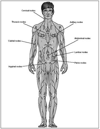
Scattered along the pathways of lymph vessels are oval or kidney bean-shaped masses of lymphatic tissue called lymph nodes, which act as filters. These nodes range in size from microscopic to just under 1 inch (2.5 centimeters) in length. The smaller lymph nodes are often called lymph nodules.
Between 500 and 1,500 lymph nodes are located in the body; most of them usually occur in clusters or chains. Principal groupings are based in the neck, armpits, chest, abdomen, pelvis, and groin. The lymph nodes in the neck, armpits, and groin are especially important because they are located where the head, arms, and legs (the extremities) meet the main part of the body (the trunk). Most injuries to the skin, which allow bacteria and other pathogens (disease-causing organisms) to enter the body, are likely to occur along the extremities. The lymph nodes at the junctions of the extremities and trunk destroy the pathogens before they reach the main part of the body and the vital organs.
In 1347, several Italian merchant ships returned to Messina on the Mediterranean island of Sicily from a trip to the Black Sea. As the ships were docking, many sailors on board were dying of a strange and hideous disease. Within days, many residents of Messina and the surrounding countryside had been infected and were dying. Within four years, the disease had spread across Western Europe and 25 million people—roughly one-third the population of Europe at the time—lay dead in its wake.
The disease was called the "Black Death" because of the black spots it produced on the skin of its victims. It is more properly known as the bubonic plague (a plague is any contagious, widespread disease). In the course of recorded history, a number of major bubonic plagues have swept across Asia and Europe. The worst, however, was the outbreak in the fourteenth century. Because so many people died, there were serious labor shortages throughout Europe. Economic growth on the continent was halted for two centuries.
Historians believe this particular plague started in China sometime in the early 1330s. As one of the world's busiest trading nations at the time, China was a prime destination for the merchant ships of Europe. But those ships brought back to the West something no one bargained for.
Bubonic plague is an infectious disease caused by Yersinia pestis , a bacteria transmitted by fleas that have fed on the blood of infected rodents, usually rats. The fleas pass on the bacteria, in turn, when they bite a human. Humans may also become infected if they have a cut or break in the skin and come in direct contact with the body fluids or tissues of infected animals or other humans.
Once inside the human body, Yersinia pestis acts quickly. Within two to five days, victims experience fever, headache, nausea, vomiting, and aching joints. The lymph nodes, especially those in the groin, then suddenly become painful and swollen. The inflamed nodes, called buboes (from which the disease gets its name), swell with pus to about the size of a chicken's egg. They soon turn black, split open, and ooze pus and blood onto the skin. The infection, rampant in the lymphatic system, quickly spreads throughout the body. Blood appears in the victim's urine and stool, and everything coming out of the body smells horrible. When death finally comes less than a week after the victim contracts the disease, it is painful.
Isolated cases of bubonic plague continue to occur in widespread regions of the world. Since the disease is caused by a bacteria, it can be treated with antibiotics. If treated quickly, the plague can almost always be cured.
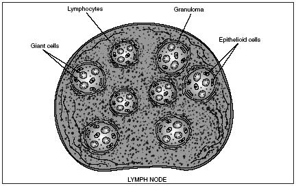
Each lymph node is enclosed in a fibrous capsule. Lymph enters the node through several small lymph vessels. Inside, bands of connective tissue divide the node into spaces known as sinuses. The specialized tissue in these sinuses harbors macrophages and lymphocytes, both of which are types of white blood cells. Macrophages engulf and destroy bacteria and other foreign substances in the lymph. Lymphocytes identify foreign substances and attempt to destroy them, also. If foreign invaders are abundant and macrophages and lymphocytes have to increase in number to defend the body against them, the lymph node often becomes swollen and tender. Once the lymph has been filtered and cleansed, it leaves the node through one or two other small lymph vessels.
Tonsils and Peyer's patches
Both tonsils and Peyer's patches are small masses of lymphatic tissue (some sources consider them specialized lymph nodes). These tissues serve to prevent infection in the body at areas where bacteria is abundant. There are five tonsils: a pair on either side of the inner wall of the throat (palatine tonsils), one near the rear opening of the nasal cavity (pharyngeal tonsil), and a pair near the base of the tongue (lingual tonsils). This "ring" around the throat helps trap and remove any bacteria or other foreign pathogens entering the throat through breathing, eating, or drinking. Peyer's patches, which resemble tonsils, are located in the small intestine. The macrophages of Peyer's patches prevent infection of the intestinal wall by destroying the bacteria always present in the moist environment of the intestine.
The spleen
The spleen is a soft, dark purple, bean-shaped organ located in the upper left side of the abdomen, just behind the bottom of the rib cage. It is the largest mass of lymphoid tissue in the body, measuring about 5 inches (12.7 centimeters) in length.
Though considered to be part of the lymphatic system, the spleen does not filter lymph (only lymph nodes do so). Instead, it filters and cleanses blood of bacteria, viruses, and other pathogens. It also destroys worn or old red blood cells. As blood flows through the spleen, macrophages lining the organ's tissues engulf and destroy both pathogens and worn red blood cells. Any remaining parts of decomposed red blood cells, such as iron, are returned to the body to be used again to form new red blood cells.
Other functions of the spleen include the production of lymphocytes, which the organ releases into the bloodstream, and blood storage. When the body demands additional blood, such as during stress or injury, the spleen contracts, forcing its stored blood into circulation.
The thymus
The thymus is a soft, flattened, pinkish-gray mass of lymphoid tissue located in the upper chest under the breastbone. In a fetus and newborn infant, the thymus is relatively large (about the size of an infant's fist). Up until about the age of puberty, the thymus continues to grow. After this point in life, it shrinks and gradually blends in with the surrounding tissue. Very little thymus tissue is found in adults.
Scientists did not fully understand the function of the thymus until the early 1960s. Only then was its role in developing body immunity discovered.
In a fetus and infant, immature or not fully developed lymphocytes are produced in the bone marrow (the spongelike material that fills the cavities inside most bones). A certain group or class of these lymphocytes travel to the thymus where thymic hormones changed them into T lymphocytes or T cells (the letter T refers to the thymus). While maturing and multiplying in the thymus, T cells are "educated" to recognize the difference between cells that belong to the body ("self") and those that are foreign ("nonself"). Each T cell is programmed to respond to a specific chemical identification marker—called an antigen—on the surface of foreign or abnormal cells. Once they are fully mature, T cells then enter the bloodstream and circulate to the spleen, lymph nodes, and other lymphatic tissue.
Lymphocytes
Lymphocytes, the primary cells of the lymphatic system, make up roughly one-fourth of all white blood cells in the body. Like other white blood cells, they are produced in the red bone marrow. Lymphocytes constantly travel throughout the body, moving through tissues or through the blood or lymph vessels.
There are two major classes of lymphocytes: T cells and B cells. As already stated, the letter T refers to the thymus, where those lymphocytes mature. The letter B refers to the bone marrow, where that group of lymphocytes mature.
About three-quarters of the circulating lymphocytes are T cells. They carry out two main defensive functions: they kill invaders and orchestrate or control the actions of other lymphocytes involved in the immune process or response. In addition, T cells recognize and destroy any abnormal body cells, such as those that have become cancerous.
Like T cells, B cells are also programmed to recognize specific antigens on foreign cells. When stimulated during an immune response (such as when foreign cells enter the body), B cells undergo a change in structure. They then produce antibodies, which are protein compounds. These compounds bind with specific antigens of foreign cells, labeling those cells for destruction.

Comment about this article, ask questions, or add new information about this topic: