
The Nervous System and the Brain - The brain
The appearance of the brain within the skull has been described as a huge gray walnut and a cauliflower. The inelegance of such descriptions is the least of many good reasons why we should be happy our brains are not exposed to public view.
Brain tissue—pinkish gray and white—is among the most delicate in our body, and the destruction of even a small part may mean lasting impairment or death. Its protection is vital and begins (if we are so fortunate) with a mat of hair on the top, back, and sides of our skull. Next comes the resilient layer of padding we call the scalp, and then the main line of defense—the rounded, bony helmet of skull.
The brain's armor does not stop with bone. Beneath are three strong, fibrous membranes called meninges that encase the brain in protective envelopes. Meninges also overlie the tissue of the spinal cord; infection or inflammation of these membranes by bacteria or viruses is called cerebrospinal meningitis .
Between two of the meninges is a region laced with veins and arteries and filled with cerebrospinal fluid . This fluid-filled space cushions the brain against sudden blows and collisions. The cerebrospinal fluid circulates not only about the brain but through the entire central nervous system. Incidentally, chemical analysis of this fluid, withdrawn by inserting a hypodermic needle between vertebrae of the spinal column (a spinal tap), can provide clues to the nature of brain and nervous system disorders.
The Cerebrum
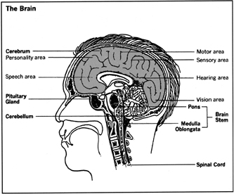
What we usually mean by “brain” is that part of the brain called the cerebrum . It is the cerebrum that permits us all our distinctly human activities—thinking, speaking, reading, writing. Only in man does the cerebrum reach such size, occupying the interior of our entire dome above the level of the eyes.
The surface of the cerebrum is wrinkled, furrowed, folded and infolded, convoluted, fissured—anything but smooth. This lavishly wrinkled outer layer of the cerebrum, about an eighth of an inch thick, is called the cerebral cortex . From its grayish color comes the term “gray matter” for brain tissue. There is a pattern among its wrinkles, marked out by wider, or deeper fissures, or furrows, running through the brain tissue. The most conspicuous fissure runs down the middle, front to back, dividing the cerebrum into two halves, the left hemisphere and the right hemisphere. The nerves from the left half of the body are served by the right hemisphere, and the right half of the body by the left hemisphere, so that damage to one side of the brain affects the other side of the body.
The Lobes of the Cerebrum
Smaller fissures crisscross the cerebrum and mark out various specific areas of function called lobes . The frontal lobes, one on the left hemisphere and one on the right in back of our eyes and extending upward behind the forehead, are perhaps the most talked about and the least understood by medical researchers. The specific functions of most other lobes in the cerebrum, such as the two occipital lobes (centers for seeing), and the temporal lobes (centers for hearing) are much better known.
The Brain Stem
The cerebrum, like a large flower obscuring part of its stalk, droops down around the brain stem . Thus, while the brain stem originates just about in the middle of our skull, it does not emerge completely from the folds of the cerebral hemispheres until it reaches the back of the neck. Then it soon merges into the spinal cord.
Associated with this portion of the brain—roughly speaking, between the cerebrum and the spinal cord—are centers that take care of the countless necessary details involved in just plain existing, and structures (such as the medulla oblongata and the pons ) that serve also as traffic control points for the billions of nerve impulses traveling to and from the cerebrum. The largest of these “lesser brains” is the cerebellum , whose two hemispheres straddle the brain stem at the back of the head.
The Cerebellum
The cerebellum is the site of balance and body and muscle coordination, allowing us, for example, to “rub the tummy and pat the head” simultaneously, or tap the foot and strum a guitar, or steer a car and operate the foot pedals. Such muscle-coordinated movements, though sometimes learned only by long repetition and practice, can become almost automatic—such as reaching for and flicking on the light switch as we move into a darkened room.
But many other activities and kinds of behavior regulated by the part of the brain below the cerebrum are more fully automatic: the control of eye movement and focusing, for example, as well as the timing of heartbeat, sleep, appetite, and metabolism; the arousal and decline of sexual drives; body temperature; the dilation and constriction of blood vessels; swallowing; and breathing. All these are mainly functions of the autonomic nervous system , as opposed to the more voluntary actions controlled by the central nervous system .
Comment about this article, ask questions, or add new information about this topic: