The Circulatory System, the Heart, and Blood - The heart at work
The structure and performance of the heart, at first glance rather complicated, assume a magnificent simplicity once we observe that this pulsating knot of hollow, intertwining muscle uses only one beat to perform two distinct pumping jobs.
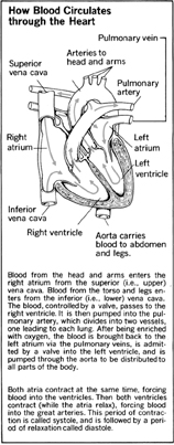
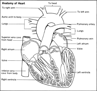
The heart has a right side (your right) and a left side (your left), divided by a tough wall of muscle called a septum . Each side has two chambers, an upper one called an atrium (or auricle) , and a lower one called a ventricle .
How the Heart Pumps the Blood
Venous blood from the body flows into the right atrium via two large veins called the superior vena cava (bringing blood from the upper body) and the inferior vena cava (bringing blood from the lower part of the body). Where the blood enters the right atrium are valves that close when the atrium chamber is full.
Then, through a kind of trapdoor valve, blood is released from the right atrium into the right ventricle. When the right ventricle is full, and its outlet valve opens, the heart as a whole contracts—that is, pumps.
To the Lungs
The blood from the right ventricle is pumped to the lungs through the pulmonary artery to pick up oxygen. The trapdoor valve between the right atrium and ventricle has meanwhile closed, and venous blood again fills the right atrium.
Having picked up oxygen in the lungs, blood enters the left atrium through the right and left pulmonary veins. (They are called veins despite the fact that they carry the most oxygen-rich blood, because they lead to the heart; just as the pulmonary artery carries the oxygen-poorest blood away from the heart, to the lungs.) Like the right atrium, the left atrium serves as a holding reservoir and, when full, releases its contents into the left ventricle. A valve between left atrium and ventricle closes, and the heart pumps.

To the Body
Blood surges through an opening valve of the left ventricle into the aorta, the major artery that marks the beginning of blood's circulation throughout the body. The left ventricle, because it has the job of pumping blood to the entire body rather than just to the lungs, is slightly larger and more muscular than the right ventricle. It is for this reason, incidentally, that the heart is commonly considered to be on our left. The organ as a whole, as noted earlier, is located at the center of the chest.
The Heart's Own Circulatory System
Heart tissue, like that of every other organ in the body, must be continually supplied with fresh, oxygen-rich blood, and used blood must be returned to the lungs for reoxygenation. The blood inside the heart cannot serve these needs. Thus the heart has its own circulation network, called coronary arteries and veins , to nourish its muscular tissues. There are two major arteries on the surface of the heart, branching and rebranching eventually into capillaries. Coronary veins then take blood back to the right atrium.
Structure of the Heart
The musculature of the heart is called cardiac muscle because it is different in appearance from the two other major types of muscle. The heart muscle is sometimes considered as one anatomical unit, called the myocardium . A tough outer layer of membranous tissue, called the pericardium , surrounds the myocardium. Lining the internal chambers and valves of the heart, on the walls of the atria and ventricles, is a tissue called the endocardium .
These tissues, like any others, are subject to infections and other disorders. An infection of the endocardium by bacteria is called bacterial endocarditis . (Disease of the valves is also called endocarditis , although it is a misnomer.) An interruption of the blood supply to the heart muscle is called a myocardial infarction , which results in the weakening or death of the portion of the myocardium whose blood supply is blocked. Fortunately, in many cases, other blood vessels may eventually take over the job of supplying the blood-starved area of heart muscle.
Heartbeat
The rate at which the heart beats is controlled by both the autonomic nervous system and by hormones of the endocrine system. The precise means by which the chambers and valves of the heart are made to work in perfect coordination are not fully understood. It is known, however, that the heart has one or more natural cardiac pacemakers that send electrical waves through the heart, causing the opening and closing of valves and muscular contraction, or pumping, of the ventricles near the normal adult rate of about 72 times per minute.
One particular electrical impulse (there may be others) originates in a small area in the upper part of the right atrium called the sinus node . Because it is definitely known that the contraction of the heart is electrically activated, tiny battery-powered devices called artificial pacemakers have been developed that can take the place of a natural pacemaker whose function has been impaired by heart injury or disease. Through electrodes implanted in heart tissue, such devices supply the correct beat for a defective heart. The bulk of the device is usually worn outside the body or is implanted just under the skin.
The fact that both ventricles give their push at the same time is very significant. It allows the entire heart muscle to rest between contractions—a rest period that adds up to a little more than half of a person's lifetime. Without this rest period, it is more than likely that our hearts would wear out considerably sooner than they do.
Blood Pressure
A physician's taking of blood pressure is based upon the difference between the heart's action at its period of momentary rest and at the moment of maximum work (the contraction or push). The split-second of maximum work, at the peak of the ventricles’ contraction, is called the systole . The split-second of peak relaxation, when blood from the atria is draining into and filling up the ventricles, is called the diastole .
Blood pressure measures the force with which blood is passing through a major artery, such as one in the arm, and this pressure varies between a higher systolic pressure, corresponding to the heart's systole, and a lower diastolic pressure, reflecting the heart's diastole, or resting phase. The device with which a physician takes your blood pressure, called a sphygmomanometer , registers these higher and lower figures in numbers equivalent to the number of millimeters the force of your arterial blood would raise a column of mercury. The higher systolic force (pressure) is given first, then the diastolic figure. For example, 125/80 is within the normal range of blood pressure. Readings that are above the normal range—and stay elevated over a period of time—indicate a person has high blood pressure, or hypertension.
Hypertension has no direct connection with nervous tension, although the two may be associated in the same person. What it does indicate is that a heart is working harder than the average heart to push blood through the system. In turn, this may indicate the presence of a circulatory problem that might eventually endanger health. See also Ch. 9, Diseases of the Circulatory System and Ch. 10, Heart Disease .


Comment about this article, ask questions, or add new information about this topic: